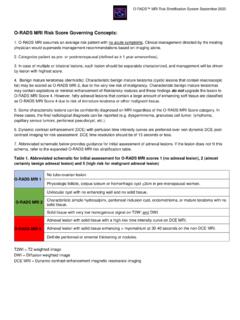
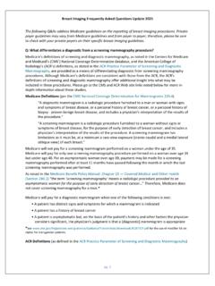
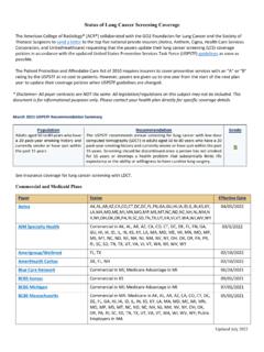
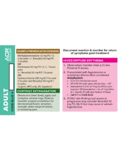
Transcription of MAMMOGRAPHY ULTRASOUND MAGNETIC RESONANCE …
1 ACR BI-RADS Atlas Fifth Edition For the complete Atlas, visit REFERENCEMAMMOGRAPHYULTRASOUNDB reast compositiona. The breasts are almost entirely fattyTissue composition (screening only)a. Homogeneous background echotexture fatb. There are scattered areas of fibroglandular densityb. Homogeneous background echotexture fibroglandularc. The breasts are heterogeneously dense, which may obscure small massesc. Heterogeneous background echotextured. The breasts are extremely dense, which lowers the sensitivity of mammographyMassesShapeOvalMassesShape Oval RoundRound IrregularIrregular MarginCircumscribedOrientation Parallel ObscuredNot parallel MicrolobulatedMargin Circumscribed IndistinctNot circumscribedSpiculated- Indistinct DensityHigh density - Angular Equal density - Microlobulated Low density - Spiculated Fat-containingEcho pattern Anechoic Calcifications Typically benignSkin Hyperechoic Vascular Complex cystic and solid Coarse or popcorn-like Hypoechoic Large rod-like Isoechoic RoundHeterogeneous Rim Posterior features No posterior features Dystrophic
2 Enhancement Milk of calciumShadowing Suture Combined pattern Suspicious morphologyAmorphous CalcificationsCalcifications in a massCoarse heterogeneous Calcifications outside of a massFine pleomorphic Intraductal calcificationsFine linear or fine-linear branching Associated featuresArchitectural distortionDistributionDiffuse Duct changesRegional Skin changesSkin thickeningGrouped Skin retractionLinear EdemaSegmental VascularityAbsentArchitectural distortionInternal vascularityAsymmetriesAsymmetry Vessels in rimGlobal asymmetry Elasticity assessmentSoftFocal asymmetry IntermediateDeveloping asymmetry HardIntramammary lymph nodeSpecial casesSimple cyst Skin lesionClustered microcysts Solitary dilated ductComplicated cyst Associated featuresSkin retraction Mass in or on skinNipple retraction Foreign body including implantsSkin thickening Lymph nodes intramammaryTrabecular thickeningLymph nodes axillaryAxillary adenopathy Vascular abnormalitiesAVMs (arteriovenous malformations/Architectural distortion pseudoaneurysms)
3 Calcifications Mondor diseaseLocation of lesionLateralityPostsurgical fluid collectionQuadrant and clock faceFat necrosisDepthDistance from the nippleMAGNETIC RESONANCE IMAGINGA mount of fibroglandular tissue (FGT)a. Almost entirely fat b. Scattered fibroglandular tissue c. Heterogeneous fibroglandular tissue d. Extreme fibroglandular tissue Associated featuresNipple retractionNipple invasionSkin retractionSkin thickeningBackground parenchymal enhancement (BPE)LevelMinimal Skin invasionDirect invasionMildInflammatory cancerModerate Axillary adenopathyMarked Pectoralis muscle invasionSymmetric or asymmetric Symmetric Chest wall invasionAsymmetric Architectural distortion FocusFat containing lesionsLymph nodesNormalMassesShape Oval AbnormalRound Fat necrosisIrregular HamartomaMargin Circumscribed Postoperative seroma/hematoma with fatNot circumscribed Location of lesionLocation- Irregular Depth- Spiculated Kinetic curve assessment Signal intensity (SI)
4 / time curve descriptionInitial phase Slow Internal enhancement characteristicsHomogeneous MediumHeterogeneous FastRim enhancement Delayed phase Persistent Dark internal septations PlateauWashoutNon-mass enhancement (NME)DistributionFocal ImplantsImplant material and lumen type Saline Linear Silicone - Intact - Ruptured Segmental RegionalMultiple regionsOther implant materialDiffuseLumen type - Single - Double - Other Internal enhancement patternsHomogeneousImplant location Retroglandular HeterogeneousRetropectoralClumpedAbnorma l implant contour Focal bulgeClustered ringIntramammary lymph nodeIntracapsular silicone findings Radial folds Skin lesionSubcapsular lineNon-enhancing findingsDuctal precontrast high signal on T1 WKeyhole sign (teardrop, noose)
5 CystLinguine signPostoperative collections (hematoma/seroma)Extracapsular silicone Breast Post-therapy skin thickening and trabecular thickeningLymph nodesWater dropletsNon-enhancing massPeri-implant fluidArchitectural distortionSignal void from foreign bodies, clips, ASSESSMENT CATEGORIESC ategory 0: MAMMOGRAPHY : Incomplete Need Additional Imaging Evaluation and/or Prior Mammograms for Comparison ULTRASOUND & MRI: Incomplete Need Additional Imaging EvaluationCategory 1: NegativeCategory 2: BenignCategory 3: Probably BenignCategory 4: SuspiciousMammography & ULTRASOUND : Category 4A: Low suspicion for malignancy Category 4B: Moderate suspicion for malignancy Category 4C: High suspicion for malignancyCategory 5: Highly Suggestive of MalignancyCategory 6: Known Biopsy-Proven


