Transcription of ManageMent of Liver abscess - API
1 469 ManageMent of Liver abscessAnita Dutta, Kishangunj, Sanjay Bandyopadhyay, KolkataINTRODUCTIONL iver is an important and vital organ of the body. This organ is subjected to numerous systemic infections viral, bacterial and parasitic and lies at the distal end of the portal circulation; it is therefore bathed with portal blood containing viruses, bacteria parasites, ova, products of digestion and other Hepatic or Liver abscesses are infectious, space-occupying lesions in the Liver ; the two most common abscesses being pyogenic and amoebic. Pyogenic Liver abscess (PLA) is a rare but potentially lethal condition, with a reported incidence of 20 per 1,00,000 hospital admissions in a western Its severity depends on the source of the infection and the underlying condition of the patient.
2 Amoebic Liver abscesses (ALA) are common in tropical regions mainly where Entamoeba histolytica is endemic and is more prevalent in individuals (mostly young males) with suppressed cell mediated both the types of hepatic abscesses, right lobe of the Liver is the most likely site of infection. The clinical presentation of both the types may be elusive with combination of fever, right upper quadrant pain and hepatomegaly with or without jaundice. PYOGENIC Liver abscess (PLA)EtiologyOver the past 50 years, there has been a major shift in the etiology of PLA. PLA may be of biliary, portal, arterial, or traumatic origin (Table 1). Ascending infection of the biliary tree secondary to obstruction is now the most identifiable cause of PLA. The etiology of biliary obstruction has some geographic differences: in Western countries this scenario is common in patients with malignant disease,4 while in Asia, gall stone disease and hepatolithiasis are more Between 15 to 55% patients in different series, no identifiable cause or source for PLA was found (hence called cryptogenic).
3 4-6 There is an increase in the median age of patients with PLA in recent years, that is mostly responsible for increased severity and mortality in spite of advancement in antibiotic ,7 Clinical presentation: Early symptoms during the onset of a PLA are non-specific and include malaise, nausea, ano-rexia and weight loss, headaches, myalgia, and arthralgia in most of the cases. These prodromal symptoms may be present for many weeks before the appearance of more specific symptoms, such as fever, chills and abdominal pain, although the pain is not always localized to the right upper quadrant. An abscess adjacent to the diaphragm may cause pleuritic type pain, cough and dyspnea, and when this presentation is associated with the above-mentioned non-specific symptoms, it can cause diagnostic difficulty.
4 Septic shock may occur in a few patients, especially in the setting of an obstructed biliary tree. 9 : 4 Medicine Update 2012 Vol. 22470 Plain abdominal and chest radiographs are usually too non-specific to be diagnostic. Ultrasound (USG) is the imaging modality used in the initial evaluation. The appearance on USG varies ac-cording to the stage of evolution of the Ini-tially the abscess is hyperechoic and indistinct, but with maturation and pus formation, it becomes hypoechoic with a distinct margin. When the pus is very thick, a fluid-containing lesion may be confused with a solid le-sion on USG. USG has a sensitivity of 75% to 95%, but has difficulty in detecting an abscess high in the dome of the right hemiliver and especially multiple small PLAs.
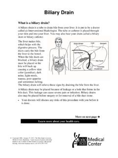
5 By showing gallstones, dilated bile ducts, and hepato-lithiasis, USG has the advantage of imaging underlying biliary tract pathology. A computed tomography (CT) scan is more accurate than USG in the differentiation of PLA from other Liver lesions and is reported to have a sensitivity of approxi-mately 95%.8 The portal venous phase using intravenous contrast material gives the best differentiation between the Liver and the abscess , with the periphery of the PLA having contrast enhancement as opposed to non-en-hancement of the central portion (Figure 1). Magnetic resonance imaging (MRI) does not seem to have any advantage over CT or USG. The spectrum of organisms that may be cultured in PLA is listed in Table 2. E. coli is cultured most frequently in Western series9,10 and Klebsiella in Asian ,7 ManageMent Of PYOGENIC Liver abscess (PLA)The principles of treatment are to Although uncommon, some patients present with peri-tonitis after free rupture of an abscess into the peritoneal cavity.
6 Diagnosis: Laboratory investigations show an elevated white cell count, anaemia, hypoalbuminemia, elevated transami-nases and alkaline phosphatase. Hyperglycemia may be the first indication that the patient is diabetic or there is loss of control because of septic process. Table 1: Common causes of pyogenic Liver abscessHepatobiliary Benign Lithiasis Cholecystitis Biliary enteric anastomosis Endoscopic biliary procedures Percutaneous biliary procedures Malignant Common bile dust Gall bladder Ampulla Head of pancreasPortal Benign Diverticulitis Anorectal suppuration Pelvic suppuration Postoperative sepsis Intestinal perforation Pancreatic abscess Appendicitis Inflammatory bowel disease Malignant Colonic cancer Gastric cancerArterial
7 Endocarditis Vascular sepsis ENT infection Dental infectionTraumatic Benign Open or closed abdominal trauma Malignant Chemoembolization Percutaneous ethanol injection or radiofrequencyCryptogenicfig. 1 : CT scan of Liver showing multiple hypodense lesions suggestive of pyogenic abscess 471 ManageMent of Liver abscess Drain the pus, Institute appropriate antibiotics, and Deal with any underlying source of infection, if present. The advances in USG and CT that provide earlier and more accurate diagnosis also have enabled these modalities to facilitate treatment through guided aspiration and drainage, shifting ManageMent away from open surgery to minimally invasive techniques. Percutaneous drainage combined with antibiotics has become the first line and mainstay of treatment for most Antibiotics: Before obtaining positive cultures from blood or pus, broad-spectrum antibiotics should be started to cover Gram-negative and Gram-positive aerobes and anaerobes.
8 Initial therapy with amoxicillin, an aminoglycosides, and metronidazole or a third generation cephalosporin and metronidazole generally covers the causative organisms most commonly found, although this regimen may be varied according to geographic differences and antimicrobial treatment policies. Initially, antibiotics should be administered parenterally, and after 2 weeks of systemic therapy, appropriate oral agents may be used for a further 4 In patients with multiple PLAs that are too small to drain, antibiotics may be the only treatment possible. In addition, efforts must be made to identify any underlying biliary obstruction, which needs to be overcome for the antibiotic therapy to procedures: Percutaneous drainage is performed under USG or CT guidance.
9 Aspiration of PLAs to confirm the diagnosis and obtain pus for culture should be accompanied by complete aspiration of the pus or insertion of a drain at the same time. Clinicians have questioned when should the primary treatment be aspiration alone and when should a percutaneous catheter drain be inserted. Percutaneous needle aspiration (PNA) group has a high success rate and a shorter hospital stay, but a large percentage requires second or third aspirations to achieve When PNA fails, catheter drainage should be performed. Primary treatment by percutaneous catheter drainage (PCD) is performed when: The pus is too thick to be aspirated The abscess is greater than 5 cm in diameter The wall is thick and non-collapsible The PLA is multi-loculated The use of PCD is not precluded by the presence of multiple abscess cavities, but this does necessitate the placement of several Although highly successful, PNA/ PCD fail in approximately 10% cases.
10 Incomplete or unsuccessful drainage may result from: Catheter too small to drain the thick pus, Position of catheter not conducive to adequate drainage, Catheter removed prematurely Thick fibrous encasing wall of PLA that is unable to col-lapse. An abscess with biliary communication has been reported to be treated as effectively by PCD as a non-communicating abscess , although the continuous output of bile leads to a prolonged period of abscess treatment: The only indication of primary surgical treatment of PLAs is in patients with an initial presentation with intraperitoneal rupture or in patients with multiple abscesses above an obstructed system that cannot be negotiated by non-operative Open operation is also indicated when there is failure of non-operative treatment and for complications of percutaneous drainage, such as bleeding or intraperitoneal leakage of pus.