Transcription of Amoebiasis - API
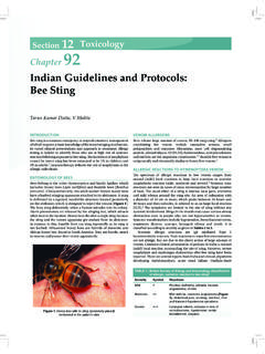
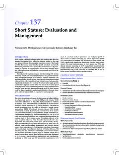
1 Be having 8,197 genes, a genome of MB size containing six isoenzymes, which help in the lysis of tissues, digestion of food material, intraluminal cellular debris and bacteria and in its penetration. Trophozoite is 20 40 in size, moves with the help of pseudopodia, formed by the flow of ectoplasm followed by the endoplasm, changing its shape. Hence, the name of Proteus is given simulating the Greek God. It has a single nucleus, food/contractile vacuole, an excretory vacuole called uroid and rudimentary mitochondria. It is also found to move at a surprising speed of 5 mm/sec (Figure 1).
2 PATHOGENESIS AND PATHOLOGYNote: Theoretically, ingestion of even one viable cyst can cause infection. Trophozoites are digested and destroyed by the gastric acid, hence, cannot cause infection even though they are ingested (Flow chart 1). The cyst divides into four initially, which divide again into eight daughter amoebae after an incubation period of 1 4 weeks, which may, however, be from few days to a year. These grow and mature into adult amoebae in about 7 10 days and stay as boarders in the large intestine, mainly the cecum and the sigmoid, feeding on intraluminal cellular debris and the bacteria.
3 The infection is usually asymptomatic. Under unfavorable conditions and as the liquid stool becomes solid during its passage down the colon, the vegetative forms become cysts and are passed in the feces. Most individuals are asymptomatic cyst shredders. AmoebiasisKVGK TilakChapter 1 DEFINITIONA moebiasis is an infection caused by Entamoeba histolytica with or without symptoms (WHO 1969). Synonyms include entamoebiasis, amoebiosis, amoebic dysentery or bloody flux. Entamoeba dispar is a harmless commensal, which is indistinguishable from E. histolytica. The other members of the group infecting humans are E.
4 Moshkovskii, E. hartmannii, E. gingivalis, Endolimax nana and Iodamoeba earliest records of bloody mucous diarrhea were found in Bhrigu Samhita (1000 BC). Asyrian and Babylonian texts (600 BC) also made a mention. Subsequently, division between amoebic and bacterial infection was made. The relationship between dysentery and liver involvement was noticed in 200 AD. Around 16th century, Amoebiasis became worldwide due to the rapid growth of trade and settlements. Accurate description of invasive and noninvasive forms of Amoebiasis was made by James Annersley in 19th century.
5 Fredrich Losch (1875) discovered amoeba in St Petersburg (Russia). Emile Brumpt (1925) suggested existence of two types of parasites, the invasive (E. histolytica) and noninvasive (E. dispar). WHO (1997) gave clear guidelines for distinguishing both the occurs worldwide, but is mostly seen in tropical and developing countries, which have bad sanitary and hygienic practices. Ten percent of world s population is estimated to be infected by the parasite (4% in USA) with an estimated annual mortality of 40,000 70,000. However, 90% of those infected are asymptomatic, 1% may develop invasive/extraintestinal Amoebiasis .
6 Spread is mostly through fecal-oral route, by ingestion of cysts and also through contaminated vegetables fertilized by feces and foods and water handled by unclean hands. Fomites and flies also have a role in the transmission. Autoinfection through improper cleaning of hands is also reported. It is uncommon in children below the age of 5 years. HIV infection peculiarly does not aggravate the illness. TAXONOMYU nicellular eukaryote-protista of Schaudin is reclassified by Cavalier Smith (1998). E. histolytica belongs to the kingdom of protozoa, subkingdom of neozoa, infrakingdom of sarcomastigota, phylum amoebezoa, and subphylum conosa.
7 E. histolytica is found to Figure 1: Structure of an amoebaSection 1 Infectious Diseases2 Infectious DiseasesSection 1inside the portal vasculature, amoebae are resistant to complement-mediated lysis. Right lobe of the liver is commonly involved, since blood draining the cecum, through a hypothetical laminar flow in portal vein, preferentially goes there. Left lobe involvement is fraught with the danger of pericardial involvement and sudden tamponade. Occasionally, small multiple abscesses occur mimicking bacterial abscesses. E. histolytica has the capacity to destroy almost all tissues of the human body, the intestinal mucosa, liver and to lesser extent the brain, skin, cartilage and even bone.
8 The virulence factors, though not known with certainty, include adhesion molecules, proteases, hemolysins, contact-dependent cytolysis, apart from its phagocytic activity. CLINICAL MANIFESTATIONSMost often, clinical manifestations are insidious and inter mittent, commencing as abdominal discomfort, bloating, irregular bowel habits, intermittent dysentery with or without blood/mucous, tenesmus with bloody mucoid diarrhea, constitutional symptoms, abdominal tenderness, toxic megacolon, and finally symptoms and signs of peritonitis secondary to perforation. Extraintestinal manifestations are primarily those of hepatic involvement.
9 These include fever, pain in right lower chest, which may be related to respiration, appetite disturbances, breathlessness, cough with or without expectoration and breathlessness, occasionally mild jaundice, rarely are secondary to severe toxemia, perforation of the bowel, toxic megacolon, rupture of the hepatic abscess into pleura, lung, peritoneum, pericardium, skin and subcutaneous tissue. Extraintes-tinal spread metastasizing in the brain and bones is uncommon. Formation of a granuloma in the bowel wall mimicking a malignant growth, the amoeboma, is also not common. Rarely, a large hepatic abscess producing obstructive jaundice can occur.
10 Fever, leukocy-tosis with elevated polymorphs, rise in hepatic enzymes and serum bilirubin are the accompaniments of the complications. DIAGNOSISHigh degree of suspicion in endemic areas is a prerequisite. Fresh liquid stool examination showing hematophagus trophozoites with Charcot-Leyden crystals is characteristic. Stool examination, preferably for three consecutive days is advocated. Presence of only cysts in asymptomatic individuals is not diagnostic, since the cysts of E. dispar, which is noninvasive and harmless are indistinguishable from those of invasive E. histolytica.