Transcription of Clinical Notification of Reportable Diseases and Conditions
1 Sincerely, Original Signed By Original Signed By Richard Baydack, PhD Carla Ens, PhD Director, Communicable Disease Control Director, Epidemiology & Surveillance Public Health and Primary Health Care Public Health and Primary Health Care Manitoba Health, Healthy Living and Seniors Manitoba Health, Healthy Living and Seniors Public Health and Primary Health Care Communicable Disease Control 4th Floor, 300 Carlton St, Winnipeg, MB R3B 3M9 T 204 788-6737 F 204 948-2040 November, 2015 Re: amebiasis Reporting and Case Investigation Reporting of amebiasis (Entamoeba histolytica) is as follows: Laboratory: All positive laboratory results for E. histolytica are Reportable to the Public Health Surveillance Unit by secure fax (204-948-3044). Health Care Professional: Probable ( Clinical ) cases of amebiasis are Reportable to the Public Health Surveillance Unit using the Clinical Notification of Reportable Diseases and Conditions form ( ) ONLY if a positive lab result is not anticipated ( , poor or no specimen taken, person has recovered).
2 Cooperation in Public Health investigations is appreciated. Regional Public Health or First Nations Inuit Health Branch (FNIHB): Once the case has been referred to Regional Public Health or FNIHB, the Communicable Disease Control Investigation Form ( ) should be completed and returned to the Public Health Surveillance Unit by secure fax (204-948-3044). 1. Case Confirmed CaseAt least one of the following: Microscopic demonstration of hematophagoustrophozoites of Entamoeba histolyticain stool orany trophozoites of Entamoeba histolyticaintissue ( , smear, aspirate, scraping, biopsy). Detection of E. histolyticaDNA by nucleic acidtesting ( , polymerase chain reaction) (PCR)in an appropriate Clinical specimen ( , ulceraspirate/biopsy). Illness clinically compatible with extra-intestinalamebiasis ( , liver abscess), accompanied bypositive serologyafor E.
3 Histolytica(1). Probable Case Microscopic demonstration of trophozoitesand/or cysts consistent with Entamoebahistolytica/disparin stool in a patient withconsistent Clinical illness from a contact of a caseof confirmed Reporting RequirementsLaboratory: All positive laboratory results are Reportable tothe Public Health Surveillance Unit (204-948-3044 secure fax). Clinical laboratories are required to submit onlyrequested residual specimens from individualswho tested positive for Entamoeba histolyticatoCadham Provincial Laboratory (CPL) withinseven days of Care Professional: Cases do not require reporting by health careprofessional as positive laboratory results will bereported to Manitoba Health by the Clinical Presentation/NaturalHistoryMost infections are asymptomatic (2, 3). Clinicalsyndromes associated with Entamoeba histolyticainfection include non-invasive intestinal tractinfection, intestinal amebiasis (amebic colitis),ameboma, and liver abscess (4).
4 Amebic diarrheawithout dysentery ( , presence of mucus andblood) is the most common disease manifestation ofinfection with E. histolyticaand the mean duration ofamebic diarrhea is three days (5). The onset ofintestinal amebiasis is often gradual with patientsreporting several weeks of symptoms (4, 6). Patientswith amoebic colitis present with bloody diarrheaand abdominal pain and tenderness (6). Disease ismore severe in those who are malnourished,pregnant women and at the extremes of age (4).Weight loss is common (4, 6), but fever is unusual(6). Symptoms may be chronic and may mimicthose of inflammatory bowel disease (4). In youngchildren with amebic dysentery, intussusceptions,perforation and peritonitis or necrotizing colitis maydevelop rapidly (5). Amebomas occur mostcommonly in the cecum and can be mistaken forcolonic carcinoma (4, 6).
5 Hematogenous dissemination may occur leading toabscesses of the liver and less commonly of the lungor brain (2). Extra-intestinal disease occurs in a smallproportion of patients (4). Amebic liver abscess, themost common extra-intestinal form of invasiveamebiasis (6, 7), is much more common in menthan in women (5). The host and parasite factorsleading to liver infection remain poorly understood(7). Symptoms of amebic liver abscess include fever,cough, and a constant, dull, aching abdominal painin the upper quadrant or epigastrium (5).Trophozoites can infect the human liver for severalmonths or years before abscesses are diagnosed (7). a Serological tests are unable to distinguish past fromcurrent infection as the level of anti-amebic antibodiesremains elevated in serum for many years (1).Communicable Disease Management ProtocolFebruary 20131 Public Health BranchCommunicable Disease Management Protocol AmebiasisAmebiasisUlceration of the skin, usually in the perianalregion, occurs rarely by direct extension fromintestinal lesions or amebic liver abscesses; penilelesions may occur in men who have sex with men(2).
6 Urinary tract problems have been described incase reports (6). 4. EtiologyAmebiasis is caused by Entamoeba histolytica, aprotozoan parasite that exists in two forms: thehardy infective cyst and the more fragile, potentiallypathogenic trophozoite (2). E. histolyticacannotgenerally be distinguished morphologically fromthe non-pathogenic E. disparand E. moshkovskii(2, 4, 5). However, hematophagous trophozoites( , that have ingested red blood cells within theircytoplasm) are pathognomonic for E. ReservoirHumans are the only known reservoir. Infectionsare usually acquired from a chronically ill orasymptomatic cyst passer (2). TransmissionTransmission usually follows ingestion of food orwater contaminated with human feces (4, 6). Fecal-oral transmission can also occur in the setting ofanal sexual practices (8) or direct rectal inoculationthrough colonic irrigation devices (4).
7 Insects,particularly flies may also act as vectors of cyst-laden feces (9). Amebic cysts are relatively chlorineresistant and can survive in moist environmentalconditions for weeks to months (2). Patients withacute amebic dysentery pose only limited danger toothers because of the absence of cysts in dysentericstools and the fragility of trophozoites (2).Persistent cyst passage may result in transmission ofE. histolyticato household contacts years later (10). OccurrenceGeneral:Entamoeba histolyticais found worldwidebut is more prevalent in people of lowersocioeconomic status who live in resource-limited countries, where the prevalence of amebic infectionmay be as high as 50% in some communities (4).The actual prevalence of E. histolyticainfection ispoorly described because much of the existing datawas collected when investigators could notdistinguish E.
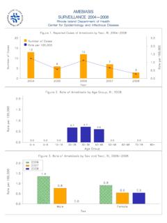
8 Histolyticafrom E. disparand (5). amebiasis is rare below the age offive years (2). Groups at increased risk of infectionin industrialized countries include immigrants fromor long-term visitors to areas with endemicinfection, institutionalized persons and men whohave sex with men (4, 11). Invasive liver disease( , liver abscess) is more common in men (8). Canada:In 2010, 637 cases of E. histolytica/disparwere reported to the National Enteric SurveillanceProgram (NESP) (12). Of the cases reported, 29were identified as being travel-associated (12).Entamoeba histolytica/disparinfections are notroutinely reported to the provincial or centralreference laboratories and are greatly under-represented in NESP (12).Manitoba:In 2011, 30 cases of amebiasis werereported to Manitoba Health. For 2000-2011inclusive, 422 cases were reported with the annualincidence varying from to per 100,000population.
9 For 2000-2011, more cases werereported in males; 266 compared to 156 in females,and the highest number of cases (214) werereported in the 20-44 year age :These data should be interpreted withcaution. Microscopic observation of stoolspecimens to detect cysts/trophozoites was thediagnostic method used to identify over 95% ofamebiasis cases reported to Manitoba cannot generally differentiate non-pathogenic Entamoeba disparor E. moshkovskiispecies. It is possible that somecases had non-pathogenic Entamoebaspeciespresent ( , E. dispar) but the gastrointestinalsymptoms were from another Incubation PeriodIncubation is usually 14 to 28 days, but rangesfrom a few days to months or years (2, 4).Communicable Disease Management ProtocolFebruary 20132 Communicable Disease Management Protocol Susceptibility and ResistanceSusceptibility to infection is general; thoseharbouring E.
10 Dispardo not develop disease (2).Re-infection is rare (2, 11). Individuals at increasedrisk of severe amebiasis , include very young or oldpersons, malnourished persons, pregnant women,and patients receiving corticosteroids (5). Period of CommunicabilityInfected patients excrete cysts intermittently,sometimes for years if untreated (4).6. DiagnosisDiagnosis is made by microscopic demonstration oftrophozoites and/or cysts in SAF-preserved fecalspecimens, and also smears, aspirates or scrapingsobtained by endoscopy/proctoscopy, aspirates ofextra-intestinal abscesses or tissue biopsies (for PCRonly). The presence of hematophagous trophozoites(containing red blood cells or their remnants)indicates invasive amebiasis caused by E. histolytica,and is the only microscopic observation thatdistinguishes E. histolyticafrom E. disparor testing (through reference laboratory) isan important or may be the sole diagnostic test forextra-intestinal amebiasis , such as amebic liverabscess, where stool examination is often sensitivity is approximately 70%.