Transcription of Microbiology: Methods for Counting Bacteria
1 MathBench- Australia Microbiology: Methods for Counting Bacteria Dec 2015 page 1 Microbiology: Methods for Counting Bacteria URL: Learning Outcomes When you have completed this module, you should be able to: Compare the advantages and disadvantages of direct counts, absorbance and plate counts as Methods for enumeration of Bacteria Recap the story Remember Matt, who went on a surfing holiday and came down with severe diarrhea? Matt, who is lying in hospital with a drip in his arm? The doctor has diagnosed cholera and is trying to work out the source. Oysters are suspected as the source of Matt s infection and samples of both the local oysters and the water around the oyster farm have been collected for testing. When the lab does the diagnostic tests, the oysters test positive for Vibrio cholerae (the bacterium that causes cholera). Now the authorities are worried that the bacterium is present in the water where the oysters are growing and they head out to collect samples.
2 They want to know how many Vibrio are present in the water. But how? Count them all? Let's get one thing clear at the outset. Given that the infective dose for cholera is about 1 million Bacteria , we are expecting to find very high numbers. Do we want to count every single one? Do we need to? Yes, I love to count: hmm, good for you but try again. No, because it takes a really long time to count a billion: y es, this is a really good reason not to count every Vibrio. So, having established that we can t count them all, what else can we do? We could sample a little of the water and a few oysters: yes, we could. We should. We will. We could give up: hmm, I don't think that's the way into a career as a MathBench- Australia Microbiology: Methods for Counting Bacteria Dec 2015 page 2 Direct count So, you have collected 5 litres of water. Back in the lab, you put a sample on a slide under a microscope, and counted everything that looked like a Vibrio.
3 That's certainly one way of quantifying the population. We'll call it .. Direct Count. Pretty easy, conceptually speaking, although it takes a bit of time to make the slide, and even more time to actually count. OK, so you've tried Counting using a Petroff Hausser chamber under the light microscope, but you can t see anything that resembles a bacterial cell. What does that mean? A less experienced lab technician might run off to tell the doctor that the water is all clear and the oysters must have been contaminated by one of the kitchen staff who handled the oysters. Not you, however, because you remember that, although this method is quick, the volume of the sample on the microscope slide is very small. This means that you will only see cells under the microscope if the concentration is high (typically more than 100,000 1 million cells per mL). If the number in the water was this high the water would actually look cloudy and no-one would have harvested those oysters!
4 ! There is another problem. Under the microscope some of the Bacteria were swimming around, but others were not moving. You wondered if that meant they were dead, but how could you tell? Light scatter (using spectrophotometer) So a direct count is a straightforward and relatively quick, but labour intensive, way to figure out how many Vibrio in the water. Wouldn't it be nice to get a machine to do the Counting for us? One way we can do this is to use a spectrophotometer. A spectrophotometer doesn't exactly count the Bacteria , but it does measure how much they interfere with a light beam , and based on that, we can use a standard curve to decide how many Bacteria there are. Then we can do the scaling up trick to get the total population size. For more details about the relationship between optical density and concentration (Beer s law; see ). This is a great method if you already have a standard curve -- perhaps you work in a lab that has been studying this particular critter for Otherwise, you may need to spend a couple of days preparing the standard curve (growing the cultures, taking the spectrophotometer readings (optical density or OD for short), comparing them to plate counts or counts under the that sort of thing).
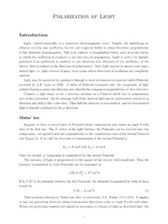
5 Here's an example of a standard curve that shows the relationship between OD measured at 650 nm and the number of Bacteria per mL: MathBench- Australia Microbiology: Methods for Counting Bacteria Dec 2015 page 3 For example, if you prepared a sample and got a spectrophotometer reading of , you would find on the x-axis and read the bacterial count off the y-axis -- about 26 million/mL Look at the graph! How about a spectrophotometer reading of (Be careful with the log scale for Bacteria !!) about 75 million/mL If your sample had about 12 million Bacteria per mL, what OD reading would you expect? OD = The dilemma of the dead cell So far we have looked at one relatively hard way (Microscopic Direct Count) and one easy way (spectrophotometer) to quantify the bacterial load in the water. But both ways share a big problem .. the problem of the dead cell. If you were Counting , say, cats .. this wouldn't really be a problem.
6 A dead cat and a live cat are generally not easily confused. If in doubt, you could poke the cat, and you would know for sure. However, bacterial cells are much less lively than cats. Yes, there is plenty of stuff going on inside, but in general you're not going to see that from the outside. Under a microscope, cells that are alive and cells that are dead look very similar most of the time. And to a standard spectrophotometer, live and dead cells look exactly alike. MathBench- Australia Microbiology: Methods for Counting Bacteria Dec 2015 page 4 Based on the dilemma of the dead cell, which would you predict? Direct count and spectrophotometer Overestimate the population: yes, these Methods overestimate, because the dead cells get counted as if they are alive Underestimate the population: the population won't be underestimated, because too many cells get counted, not too few. Get the population just right: the population will not be accurately estimated, both Methods count too many cells.
7 If we care about getting the number of live cells correct, we need a better Viable plate count So we can't poke a cell to see if it's alive -- how else can we decide if a bacterial cell is alive? What do live cells do that dead cells don't? Well, one big thing is, live cells grow and reproduce. And they do it fairly quickly (remember, every half hour or so in the case of vibrio Bacteria in the lab). So if we were to take a batch of cells and wait a day or so, we would soon know if they were alive or not. Let's refine this idea a little, by taking a look at a day in the life of a microbe. Out in the real world (like in an oyster, in water or your bloodstream), Bacteria are limited by temperature and nutrients. In the lab, we're going to put the Bacteria on a medium, which will do two things: 1. take away limiting factors by providing the nutrients and growth conditions that promote growth, and 2. fix each individual cell in one place, so it doesn't get moved around.
8 So, let s watch a single bacterial cell, conveniently named Minnie, sitting in the middle of a batch of medium. The medium has every creature comfort that Minnie could want, and she pulls in nutrients as fast as she can. Soon she s doubled in size and she divides in two, creating little Minnies 2 and 3. These two also pull in nutrients as fast as they can, and a half hour later, they too are ready to divide, and they do. Minnies 4, 5, 6 and 7 keep following the same pattern. MathBench- Australia Microbiology: Methods for Counting Bacteria Dec 2015 page 5 time: 0 min time: 30 min time: 1 hr time: 1 hr 30 min Luckily, none of the Minnies move around much. So, they re all stuck sitting in a little pile, but they don t mind, as long as there are plenty of nutrients to go around. And pretty soon we have a pile consisting of Minnies 8 through 15, all descendents of Minnie the First. And so on. Eventually (after about 24 hours) this pile will be big enough for one of us human beings to see without even using a microscope.
9 This tiny but visible pile is called a colony, and the original Minnie was the original colony-forming unit, or CFU. Of course Minnie was probably not the only CFU around. Over on the other side of the agar, Ginnie was sitting around, minding her own business, enjoying the warmth and nutrients, and growing at the same rate. So while a few million descendants of Minnie formed a colony on one side of the plate, a few million descendants of Ginnie form a colony of the other side. And so on, one colony for each original colony forming a unit (i .e . bacterial cell or clump of cells). Comparison of Methods We've talked about 3 Methods so far: just plain Counting under a microscope, using a spectrophotometer to do the dirty work, or growing Bacteria in a dish to count how many piles of descendants (colonies) they produce. There is another way that we could (theoretically) figure out how many cells there are: we could gather them all in one place and weigh them.
10 This would be the biomass method. Biomass is an important way of measuring populations in the lab, but in this case it would not help us -- we could do it but it would not tell us if the cells were dead or alive, or distinguish between particles ( s oil particles) in the water and Bacteria . So, let's compare the 3 practical ways we have of measuring the number of cells: MathBench- Australia Microbiology: Methods for Counting Bacteria Dec 2015 page 6 Direct Count Spectrophotometer Viable Plate Count Which one requires the least time (for example, the boss wants the answer before lunch)? NOT TOO BAD - Counting can be time-consuming, but the answer could be available before lunch. FASTEST - but only if you already have the calibration curve!!! SLOWEST - we have to wait an entire day for the cells to grow. But the oysters are off the menu till this is sorted, so we can wait Which one requires the least effort (think lab technician pain)?