Transcription of MODULE 6: RESPIRATORY AND DIGESTIVE SYSTEM …
1 1 MODULE 6: RESPIRATORY AND DIGESTIVE SYSTEM ANATOMY The RESPIRATORY SYSTEM includes the structures of the upper and lower RESPIRATORY tract (or from the mouth to the alveoli). The DIGESTIVE SYSTEM includes structure from the mouth to the anus. The branching point for these two systems is in the larynx where one conduit continues into the trachea past the vocal cords and the other continues down the esophagus into the stomach. The tables below includes a list of terms that you will be expected to identify on pictures, models and images. There will likely be images on the exam that you have not seen or studied before, so it is important for you to learn anatomical structures by their characteristics and avoid just memorizing the pictures you have available.
2 List of Terms Spend as much time as you need reviewing cardiovascular anatomy. Practice identifying all of the structures listed in the tables below. Use your online resources, open lab, and any other tool that you have to become confident in your identification skills. Even though the practice exam will be multiple choice, the real lab exams will ask you to identify and then write in (Fill in the Blank) the correct term for your identification. The tables below include a comprehensive list of all the terms from this section that we would consider asking about on an exam.
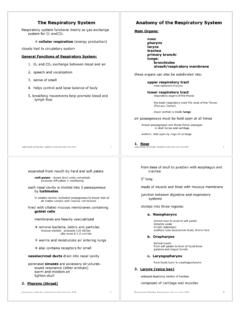
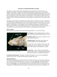
3 2 LIST OF TERMS FOR THE RESPIRATORY SYSTEM Upper RESPIRATORY Tract Nasal Cavity External Nares (Nostrils) Vestibule Nasal Septum Hard Palate Conchae Superior Conchae Middle Conchae Inferior Conchae Meatus Superior Meatus Middle Meatus Inferior Meatus Nasolacrimal Duct Choana (Internal Nares) Paranasal Sinuses Frontal Sinus Sphenoid Sinus Most folks are quite familiar with the Nostrils. Those two holes on the inferior end of our nose are hard to ignore. Just behind the nostrils is the Vestibule. This area can be hard to picture as we don t see it on the outside of the face.
4 It is circled in the picture below and to the left. The Nasal Septum is a wall of bone and cartilage that separates the two nostrils from each other. Continuing back to the posterior end of the nostrils we reach a point where the nostrils join just before the nasal cavity dips inferiorly to become the back of the throat. This point is called the Choana and is in the picture below and to the right. The Conchae are curled bone shelves located on the lateral nasal cavity in each nostril. A Meatus is the valley separating each conchae. The Nasolacrimal Duct is a passage way for water on the eyes to enter the nose.
5 The Paranasal Sinuses are hollow cavities in bones surrounding the nasal cavity. 3 Pharynx Nasopharynx Pharyngeal Tonsil (Adenoid) Soft Palate Oropharynx Fauces Palatine Tonsils Lingual Tonsils Uvula Laryngopharynx The Pharyngeal Tonsils are also called the Adenoids. This is a mass of lymphatic tissue located posterior to the nasal cavity. Basically, the adenoids are where the nose blends into the throat (See picture below). The Palatine Tonsils can be seen on the left and right sides of the throat in an anterior view through the mouth.
6 The Lingual Tonsils are rounded masses on the posterior region of the tongue (See picture below). The Fauces are the archway formed by the tongue, palatine tonsils and the soft palate. The Uvula (sometimes called the palatine uvula) is a cone shaped projection from the posterior edge of the soft palate. It functions in the production of some sounds in human speech. The Laryngopharynx is the bottom part of the throat that connects to the esophagus 4 Lower RESPIRATORY Tract Hyoid Bone The Hyoid Bone (sometimes called the lingual bone) is a horseshoe shaped bone in the anterior midline of the neck.
7 Unlike other bones this bone sits alone without any adjacent attachments to other bones. It functions in speech and swallowing. Larynx Thyroid Cartilage Cricoid Cartilage Epiglottis Arytenoid Cartilage Vestibular Folds (false vocal cords) Vocal Folds (true vocal cords) There are 9 different cartilage structures that make up the larynx. The Thyroid Cartilage is the largest. Two plate like layers form a point anteriorly that has been called the adams apple. The Cricoid Cartilage refers to a ring cricoid is Greek for ring shaped . It is the only complete ring of cartilage in the trachea.
8 The Epiglottis is a flap of elastic cartilage that covers the entrance to the larynyx while swallowing. The Vestibular Folds (sometimes called the false vocal cords) are two thick folds of tissue covered with mucus membrane. They sit just superficial to the vocal cords (see picture to left). They can constrict and close the opening to the larynx. The Vocal Folds are thin mucus membranes stretched just deep to the vestibular folds. They vibrate when they are brought close together and form sounds that become speech. The Artytenoid Cartilage are a pair of pyramid shaped structures that the vocal folds attach to.
9 Movement of the arytenoid cartilage aids in the fine movement adjustments of the vocal folds. The Trachea extends below the larynx and is composed of cartilage rings. Although the rings are not complete. The trachea is often referred to as the wind pipe . Once the trachea branches into to conduits, we call the conduits Primary or Main Bronchi. The next branching creates bronchi referred to as Secondary or Lobar Bronchi. The next branching after this yields Tertiary or Segmental Bronchi. Branching after the tertiary bronchi produces air conduits that have no cartilage on them.
10 They are tubes covered with smooth muscle and their diameters can be regulated. These tubes are collectively referred to as Bronchioles. Terminal Bronchioles do not branch anymore but dead end in a cluster of sac structures called Alveoli. There may be a small segment of terminal bronchiole that expresses some alveoli (before the main cluster). This segment is called the RESPIRATORY Bronchiole. RESPIRATORY Tree Trachea Primary (Main) Bronchi Secondary (Lobar) Bronchi Tertiary (Segmental) Bronchi Bronchioles Terminal Bronchioles RESPIRATORY Bronchioles Alveoli 5 Lungs Hilum Right Lung Superior Lobe Horizontal Fissure Middle Lobe Oblique Fissure Inferior Lobe Left Lung Superior Lobe Oblique Fissure Inferior Lobe The Hilum refers to the opening into the lung.