Transcription of MORPHOLOGY AND Notes CLASSIFICATION OF BACTERIA
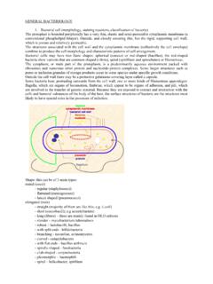
1 1 MORPHOLOGY and CLASSIFICATION of BacteriaMICROBIOLOGYMODULEM icrobiologyNotes1 MORPHOLOGY ANDCLASSIFICATION OF INTRODUCTIONM icroorganisms are a heterogeneous group of several distinct classes of livingbeings. Based on the difference in cellular organization and biochemistry, thekingdom protista has been divided into two groups namely prokaryotes andeukaryotes. BACTERIA and blue-green algae are prokaryotes, while fungi, otheralgae, slime moulds and protozoa are eukaryotes. BACTERIA are prokaryoticmicroorganisms that do not contain chlorophyll. They are unicellular and do notshow true branching, except in higher BACTERIA like reading this lesson, you will be able to.
2 Zdescribe the structure of Prokaryotic and Eukaryotic cellzexplain the size of bacteriazclassify BACTERIA based on the shape and arrangementszdescribe the structure of bacterial cell wallzdescribe the phases of Growth curvezexplain the factors affecting the growth of PROKARYOTESThe prokaryotic cells have the following characteristics such aszNo organelles, all the action takes place in the cytosol or cytoplasmicmembraneMICROBIOLOGYMODULEM orphology and CLASSIFICATION of BacteriaMicrobiology 2 NoteszMost BACTERIA possess peptidoglycan, a unique polymer that makes itssynthesis a good target for antibioticszProtein synthesis takes place in the cytosol with structurally differentribosome sFig.
3 : Prokaryote CellFig. : Eukaryote CellDifference between Prokaryotic and Eukaryotic CellsCharacterNucleusMembrane-boundorgan ellesChromosome (DNA)ProkaryotesAbsent. No nuclearenvelopeAbsentSingle coiled chromosomein cytoplasm nucleoid region in association with histone-like proteinsEukaryotesPresent with nuclearenvelope and nucleolusPresent. Includesmitochondria, chloroplasts(plants), lysosomesMultiple linearchromosomes with histoneproteins 3 MORPHOLOGY and CLASSIFICATION of BACTERIAThe major characteristics of BACTERIA are based on their size, shape SizeThe unit of measurement used in bacteriology is the micron (micrometer)1 micron ( ) or micrometer ( m) one thousandth of amillimeter1 millimicron (m ) or nanometer (nm) one thousandth of a micronor one millionth of amillimeter1 Angstrom unit ( )
4 One tenth of a nanometerThe limit of resolution with the unaided eye is about 200 microns. BACTERIA aresmaller which can be visualized only under magnification. BACTERIA of medicalimportance generally measure m in diameter and about 3-5 m wallMitotic divisionRibosomesFlagellaCytoplasmic membranelipidsMitochondriaLysosomesGolgi apparatusEndoplasmic ReticulumEubacteria have a cell wallof peptidoglycan Archaeahave cell walls ofpseudomureinAbsent70S. Free in cytoplasmwhen present consist ofprotein flagellinEubacteria= Fatty acidsjoined to glycerol by esterlinkageArchaea=Hydrocarbons joined toglycerol by ether linkageAbsentAbsentAbsentAbsentNo cell wall in animalcellsPlant cell walls =celluloseFungal cell walls =chitinPresent80S.
5 Both free in cytoplasmand attached to in mitochondriaand chloroplastsconsist of 9+2 arrangementof microtubulesFatty acids joined toglycerol by ester linkagePresentPresentPresentPresentMICRO BIOLOGYMODULEM orphology and CLASSIFICATION of BacteriaMicrobiology MicroscopyThe morphological study of BACTERIA requires the use of microscopes. Microscopyhas come a long way since Leeuwenhoek first observed BACTERIA using hand-ground types of microscope are(i) Light or optical microscope(ii) Phase contrast microscope(iii) Dark field/ Dark ground microscope(iv) Electron microscopeLight or optical microscopeThey are of two types namely Simple and Compund MicroscopezSimple Microscope consists of a single lens.
6 A hand lens is an example ofa simple Microscope consists of two or more lenses in series. The imageformed by the first lens is further magnified by another may be examined under the compound microscope, either in the livingstate or after fixation and staining. Examination of wet films or hanging dropsindicates the shape, arrangements, motility and approximately size of the due to lack of contrast details cannot be contrast microscopeThis imposes the contrast and makes evident the structure within the cells thatdiffer in thickness or refractive index. The difference in the refractive indexbetween BACTERIA cells and the surrounding medium makes them clearly , by a fraction of a wavelength, of the rays of light that pass throughthe object, compared to the rays passing through the surrounding medium,produces phase difference between the two types of field / Dark ground microscopeAnother method of improving the contrast is the dark field microscope in whichreflected light is used instead of the transmitted light used in the ordinalmicroscope.
7 The contrast gives an illusion of increased resolution, so that veryslender organisms such as spirochete, not visible under ordinary illumination,can be clearly seen under the dark field microscope. 5 MORPHOLOGY and CLASSIFICATION of BacteriaMICROBIOLOGYMODULEM icrobiologyNotesElectron MicroscopeBeams of electron are used instead of beam of light, used in light object which is held in the path of beam scatters the electrons and producesan image which is focused on a fluorescent viewing screen. Gas moleculesscatter electron, therefore it is necessary to examine the object in a QUESTIONS the microscope(a) reflected contrast microscope(b) electron field microscope(c) light microscope(d) refractive Stained PreparationsLive BACTERIA do not show the structural detail under the light microscope dueto lack of contrast.
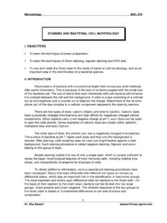
8 Hence staining techniques are used to produce colourcontrast. Routine methods of staining of BACTERIA involve dying and fixingsmears procedures that kill them. BACTERIA have an affinity to basic dyes dueto acidic nature of their protoplasm. The commonly used staining techniques areSimple StainsDyes such as methylene blue or basic fuchsin are used for simple staining. Theyprovide colour contrast, but impart the same colour to all StainingBacteria are mixed with dyes such as Indian ink or nigrosin that provide auniformly coloured background against which the unstained BACTERIA stand outin contrast.
9 Very slender BACTERIA like spirochetes that cannot be demonstratedby simple staining methods can be viewed by negative MethodsCells and structures too thin to be seen under ordinary microscope may berendered visible if they are impregnated with silver on the surface. These areused for demonstration of spirochetes and bacterial and CLASSIFICATION of BacteriaMicrobiology 6 NotesDifferential StainsThese stains impart different colours to different BACTERIA or bacterial structures,the two most widely used differential stains are the Gram stain and Acid faststain.
10 The gram stain was devised by histologist Christian Gram as a method ofstaining BACTERIA in positive cells are simpler chemical structure with a acidic protoplasm. Ithas a thick peptidoglycan layer. Teichoic acids are intertwined among thepeptidoglycan and the teichoic acids are the major surface antigen determinantsGram negative cells are more complex, they are rich in lipids. The membraneis bilayered as phospholipids, proteins and lipopolysaccharide. Lipopoly-saccharides (LPS) are also known as endotoxin. Gram negative cells have apeptidoglycan layer which is thin and formed by just one or two molecules.