Transcription of MSK MRI PROTOCOLS - bonepit.com
1 MSK MRI protocol OVERVIEW Page 1 of 123 MSK MRI PROTOCOLS March 2010 TABLE OF CONTENTS UNSUPERVISED PROTOCOLS SHOULDER (ROUTINE) ..2 SHOULDER + PROXIMAL biceps ..6 ELBOW (ROUTINE) ..10 ELBOW + distal biceps TENDON ..13 ELBOW + distal TRICEPS TENDON ..17 WRIST (ROUTINE) ..20 WRIST + DRUJ INSTABILITY ..23 THUMB ..26 HIP HIGH RESOLUTION ..29 HIP AVN, OA, HIP FRACTURE ..33 PELVIS (ALL BONY PELVIS) ..36 SACROILIAC JOINTS ..38 SACRUM/COCCYX AND SACROILIAC JOINTS ..40 QUADRICEPS TENDON/MUSCLE ..43 HAMSTRING TENDONS/MUSCLES ..47 KNEE (ROUTINE) ..50 CALF/TIBIA STRESS FRACTURE protocol ..53 ANKLE (ROUTINE) ..57 ANKLE (FLEXOR AND PERONEAL TENDONS) ..60 ACHILLES TENDON ..63 MORTON S NEUROMA/FOREFOOT (WITH GAD) ..65 TEMPOROMANDIBULAR JOINTS protocol ..68 SPONDYLOARTHROPATHY protocol .
2 71 SUPERVISED PROTOCOLS SHOULDER ARTHROGRAM (POST ONLY)..75 SCAPULA ..80 PECTORALIS MAJOR ..82 STERNOCLAVICUALR JOINTS ..87 ELBOW ARTHROGRAM (PRE AND POST) ..89 WRIST ARTHROGRAM (PRE AND POST) ..92 HAND ..95 HIP ARTHROGRAM (PRE AND POST) ..98 LUMBOSACRAL PLEXUS ..105 SPORTS HERNIA/ PUBALGIA/ OSTEITIS PUBIS ..107 KNEE ARTHROGRAM (POST ONLY) ..110 ANKLE ARTHROGRAM (PRE AND POST) ..112 MIDFOOT/FOREFOOT ..117 LIS FRANC INJURY ..120 MISCELLANEOUS TECHNIQUES FOR REDUCING METAL ARTIFACT ON MR IMAGING ..123 MSK MRI protocol OVERVIEW Page 2 of 123 MSK MRI PROTOCOLS March 2010 SHOULDER (ROUTINE) GENERAL COMMENTS - Supraspinatus tendon is what you use to plan the coronal sequence for a routine shoulder - Glenohumeral joint is what you use to plan the coronal and sagittal sequences for a post arthrogram shoulder COIL - Shoulder coil - Large 4 channel flex coil for larger patient (make certain that coverage has enough signal for the AC joint) - Body matrix coil for an extremely large patient POSITIONING - Supine - Try to have shoulder neutral (external rotation is fine) - Try to limit superior or inferior positioning of shoulder when compared to the chest (IF THE SHOULDER IS MARKEDLY ANGLED, YOU CAN ANGLE THE AXIAL IMAGES PERPENDICULAR TO THE GLENOHUMERAL JOINT)
3 - Try to place shoulder as close to isocenter in the bore of the magnet - Place a sponge at the elbow and one supporting the hand and strap the arm in place SEQUENCES, AND WHAT IS MOST IMPORTANT TO REPEAT - Cor T2 FS is most important sequence to repeat if motion - Axial PD FS is next most important sequence 1. AX T1 2. AX PD FS 3. COR T2 FS 4. COR T2 5. SAG T2 FS 6. SAG T2 SEQUENCES LOC 3 PLANE LOC MSK MRI protocol OVERVIEW Page 3 of 123 MSK MRI PROTOCOLS March 2010 1. AXIAL T1 AND PD FS - Use coronal LOC and plane is straight horizontal (IF THE SHOULDER IS MARKEDLY ANGLED, YOU CAN ANGLE THE AXIAL IMAGES PERPENDICULAR TO THE GLENOHUMERAL JOINT) - Cover from top of AC joint down and try to cover to the inferior portion of the glenohumeral joint axillary pouch - No Sat Band 2.
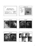
4 COR T2 AND T2 FS - Use axial T1 or PD FS sequence to orient the plane along the supraspinatus tendon (first image) - If the supraspinatus tendon is not well seen, you can either use the teres minor tendon (second image) or the glenohumeral joint MSK MRI protocol OVERVIEW Page 4 of 123 MSK MRI PROTOCOLS March 2010 - Cover from anterior portion of coracoid process to 1 slice posterior to the humeral head. - Oblique Sat band over chest 3. SAG T2 FS - Perpendicular to Coronal sequence - Angle approximately parallel to GH joint on the Cor T2 sequence (use Glenoid articulating surface to angle) - Cover from 1 slice out of humeral head to as far medial as slices allow - Oblique Sat band over chest MSK MRI protocol OVERVIEW Page 5 of 123 MSK MRI PROTOCOLS March 2010 4.
5 SAG T2 - The slices are thicker than the SAG T2 FS (to cover more of the muscle bellies) - Oblique Sat band over chest - Cover from 1 slice out of humeral head to as far medial as the slices go (to approx. the medial portion of the coracoid process) MSK MRI protocol OVERVIEW Page 6 of 123 MSK MRI PROTOCOLS March 2010 SHOULDER + PROXIMAL biceps GENERAL COMMENTS - Covers shoulder and down to mid-to- distal humerus - Can t use shoulder coil because it does not work with the body matrix coil COIL - Large 4 channel flex coil over shoulder - Body matrix coil over upper arm, minimally overlapped with flex coil - Use both coils at the same time only for the SAG T2 POSITIONING - Supine - Try to have shoulder neutral (external rotation is fine) - Try to limit superior or inferior positioning of shoulder when compared to the chest - Try to place shoulder as close to isocenter in the bore of the magnet - Place a sponge at the elbow and one supporting the hand and strap the arm in place SEQUENCE ORDER SHOULDER 1.
6 AX T1 2. AX PD FS 3. COR T2 FS 4. COR T2 5. SAG T2 FS SHOULDER AND UPPER ARM 7. SAG T2 UPPER ARM 8. AX T1 9. STIR SEQUENCES LOC (BOTH COILS ON FOR LOC) 3 PLANE LOC MSK MRI protocol OVERVIEW Page 7 of 123 MSK MRI PROTOCOLS March 2010 1. AXIAL T1 AND PD FS - Use coronal LOC and axial plane is straight horizontal (don t angle) - Cover from top of AC joint down and try to cover to the inferior portion of the glenohumeral joint axillary pouch - No Sat Band 2. COR T2 AND T2 FS - Use axial T1 or PD FS sequence to orient the plane along the supraspinatus tendon. - If the supraspinatus tendon is not well seen, you can either use the teres minor tendon or the scapular body - Cover from anterior portion of coracoid process to 1 slice posterior to the humeral head. - Oblique Sat band over chest MSK MRI protocol OVERVIEW Page 8 of 123 MSK MRI PROTOCOLS March 2010 3.
7 SAG T2 FS - Perpendicular to Coronal sequence - Angle parallel to GH joint or humeral shaft on the Cor T2 sequence - Cover from 1 slice out of humeral head to as far medial as slices allow - Oblique Sat band over chest MSK MRI protocol OVERVIEW Page 9 of 123 MSK MRI PROTOCOLS March 2010 4. SAG T2 - The FOV is bigger, the slices are thicker, and there are more slices ( to cover most of the muscles of the upper arm) - Try and use same alignment as for the SAG T2 FS - Oblique Sat band over chest 5. AXIAL T1 - Overlap 1-2 slices with Axials of Shoulder - Cover down as far as slices allow - Sat band above 6. AXIAL STIR - Overlap 1-2 slices with Axials of Shoulder - Cover down as far as slices allow - Parallel Sat bands above and below MSK MRI protocol OVERVIEW Page 10 of 123 MSK MRI PROTOCOLS March 2010 ELBOW (ROUTINE) COIL - Use 4 Channel Flex coil - Make sure the coil is centered at the olecranon POSITIONING - Supine (or if a large patient in the Superman position) - Try to have elbow fully extended - Try to have hand supinated (palm up and put sandbag on hand) - Elevate elbow with a sponge to isocenter (if supine) - Sponge and strap elbow in place SEQUENCES, AND WHAT IS MOST IMPORTANT TO REPEAT - Axial PD and COR STIR sequences are the most important to repeat 1.
8 COR STIR 2. COR T1 3. AX PD 4. AX STIR 5. SAG PD FS SEQUENCES AX LOC 3 PLANE LOC 1. CORONAL T1 AND STIR SEQUENCES - Use axial LOC to angle parallel to anterior portions of the capitellum and trochlea (or parallel to humeral epicondyles) MSK MRI protocol OVERVIEW Page 11 of 123 MSK MRI PROTOCOLS March 2010 - Use sagittal LOC to angle parallel to humerus/radius/ulnar plane, but closer to plane of radius if minimally flexed (if markedly flexed elbow, then angle between anterior humerus and the radius) - Cover from back of the olecranon to at least 1 slice anterior to radial head 2. AXIAL PD AND STIR SEQUENCES - Perpendicular to Coronal - Use COR T1 to angle parallel to elbow joint (parallel to capitellum and trochlea) - Cover from 1 slice distal to radial tuberosity up as far as the slices go - Parallel Sat Bands (above and below) MSK MRI protocol OVERVIEW Page 12 of 123 MSK MRI PROTOCOLS March 2010 3.
9 SAGITTAL PD FS SEQUENCE - Perpendicular to both Coronal and Axial sequences - Cover 1 slice outside of both humeral epicondyles - No Sat Band MSK MRI protocol OVERVIEW Page 13 of 123 MSK MRI PROTOCOLS March 2010 ELBOW + distal biceps TENDON GENERAL COMMENTS - Larger FOV than normal elbow (to cover more of the muscle) - Covers more anteriorly (covers biceps muscle) - Need to cover 1 slice distal to radial tuberosity COIL - Use Body Matrix Coil (to cover higher) - Make sure the coil covers from the radial tuberosity to as high on the humerus as the coil goes POSITIONING - Supine (or if a large patient in the Superman position) - Try to have elbow fully extended - Try to have hand supinated - Elevate elbow with a sponge to isocenter (if supine) - Sponge and strap elbow in place SEQUENCES, AND WHAT IS MOST IMPORTANT TO REPEAT - Axial PD and COR STIR sequences are the most important to repeat - Then repeat SAG PD FS 1.
10 COR STIR 2. COR T1 3. AX PD 4. AX STIR 5. SAG PD FS Reposition 6. FABS PD FS SEQUENCES AX LOC 3 PLANE LOC MSK MRI protocol OVERVIEW Page 14 of 123 MSK MRI PROTOCOLS March 2010 1. CORONAL T1 AND STIR SEQUENCES - Use axial LOC to angle parallel to anterior portions of the capitellum and trochlea (or parallel to humeral epicondyles) - Use sagittal LOC to angle parallel to humerus/radius/ulna plane (if flexed then angle more parallel to humerus) - Cover from anterior muscles back to at least mid-portion of olecranon - No Sat Bands 2. AXIAL PD AND STIR SEQUENCES - Perpendicular to Coronal - Use COR T1 to angle parallel to elbow joint (parallel to capitellum and trochlea) - Cover from 1 slice distal to radial tuberosity and up as far as the slices go - Parallel Sat Bands - Can do a second PD and STIR stack higher up biceps muscle if the abnormal muscle is not all covered (Or slightly thicker slices if the extra coverage is not that much) MSK MRI protocol OVERVIEW Page 15 of 123 MSK MRI PROTOCOLS March 2010 3.