Transcription of Shoulder Impingment - bonepit.com
1 Shoulder ImpingmentJong Liu05/18/06 What is it? Rotator cuff impingement syndrome is a clinical diagnosis that is caused by mechanical impingement of the rotator cuff by its surrounding structures. Patients with impingement syndromes may present with various signs and symptoms on physical examination depending on the degree of pathology and the structures involved. Subacromial Impingement Narrowing of the space between the humeral head and the coracoacromial arch (supraspinatus outlet); Causing entrapment of the supraspinatus tendon and subacromial bursa.
2 Repeated trauma to these structures will lead to tendon degeneration/tear and bursitis. Patients complain of pain and tenderness over anterior or anterolateral aspect of the Shoulder joint. Subacromial Impingement Neer proposed that 95% of rotator cuff tears are due to chronic impingement between the humeral head and the coracoacrominal Impingement Stage 1 disease consists of edema and hemorrhage of the tendon due to occupational or athletic overuse, and is reversible under conservative Impingement Stage 2 disease shows progressive inflammatory changes of the rotator cuff tendons and the subacromial-subdeltoid bursa.
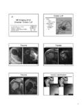
3 And can be treated by removing the bursa and dividing the coracoacromial ligament after failed conservative Impingement Stage 3 disease manifests as partial or complete tears of the rotator cuff and secondary bony changes at the anterior acromion, the greater tuberosity or the acromioclavicular Impingement abnormal acromial shape or position; subacromial enthesophytes; os acromiale; thickened coracoacromial ligament; acromioclavicular joint undersurface osteophytes. Subacromial Impingement Morrison and Bigliani described three types of acromion based on dried cadaver specimens and conventional outlet view radiographs.
4 Type 1 acromion has a flat undersurface and is considered the physiologic shape. Type 2 acromion has a curved undersurface. Type 3 acromion has a hooked undersurface. Subacromial Impingement Both type 2 and 3 acromion are considered abnormal variants that predispose individuals to impingement of supraspinatus beneath the acromion, and increase the likelihood of developing rotator cuff tear. Type IType IIType IIIType III AcromionSubacromial EnthesophyteLow lying acromionAC Joint Undersurface OsteophyteThickened Coracoacromial ligamentOs AcromialeSubcoracoid Impingement The coracoid process may cause anterior impingement when the coracohumeral distance is decreased.
5 This distance must be large enough to accommodate the articular cartilage of the humerus, the subscapularis tendon, the subscapularis bursa and the rotator interval tissue, and portions of the insertions of the coracoacromial ligament and the conjoint tendon. Subcoracoid Impingement Gerber s study in normal subjects with conventional CT of the Shoulder demonstrates average distance between medially rotated humeral head (the lesser tuberosity) and the coracoid tip is mm. Forward flexion combined with medial rotation reduced the coracohumeral distance to an average of mm (30).
6 A coracohumeral space of less than 6 mm was considered diagnostic of subcoracoid ImpingementSubcoracoid Impingement anatomic abnormality of the coracoid process such as longitudinally or laterally displaced coracoid process, or developmental enlargement of the coracoid process. surgical procedures involving the coracoid process, such as bone block procedures for anterior instability of the Shoulder , posterior glenoid neck osteotomies for posterior instability of the Shoulder , and acromionectomies for rotator cuff tears.
7 Fractures of the lesser tuberosity or the coracoid process, and subsequent malunion that leads to decreased subcoracoid space. lesions in the subcoracoid space such as ganglions, calcifications, and amyloid deposits. Subcoracoid Impingement Most patients complain of pain and tenderness in the anterior aspect of the Shoulder , which is exacerbated by various degrees of flexion, adduction, and rotation. The pain is thought to be caused by impingement of the subscapularis tendon between the lesser tuberosity and coracoid process.
8 Modified Kennedy-Hawkins SignTest performed with the arm flexed 90 , adducted 10 , and internally rotatedSubcoracoid Impingement MR axial and oblique sagittal images are used to evaluate the coracohumeral space and subcoracoid impingement. Subscapularis tendon partial or full thickness tear and biceps tendon instability has been reported in patients with clinical diagnosis of subcoracoid ImpingementSubcoracoid ImpingementSubcoracoid ImpingementSecondary Extrinsic Impingment In patients with symptoms of secondary extrinsic impingement, the coracoacromial outlet is usually normal.
9 Overhead-throwing athletes can develop glenohumeral joint instability secondary to fatigue and overloading of the rotator cuff muscles caused by chronic microtrauma and weakening of the anterior capsule. This instability will cause abnormal superior translation of the humeral head and lead to dynamic narrowing of the coracoacromial outlet. Instability can also occur in the scapulothoracic joint, and cause abnormal scapular motion and result in dynaminc narrowing of the coracoacromial outlet. Secondary Extrinsic Impingment MR images will show undersurface degeneration and partial tears of the rotator cuff tendons.
10 Labral abnormality is also described in patients with secondary extrinsic impingement. Posterosuperior glenoid impingement Posterosuperior glenoid impingement syndrome was first described by Walch et al in athletes who participate in recurrent overhead activities, such as throwing, tennis playing, and swimming. Posterosuperior glenoid impingement During the late cocking phase of throwing motion, the arm is maximally abducted and maximally externally rotated. This extreme ABER position will cause contact between the undersurface fibers on the supraspinatus and infraspinatus and posterosuperior glenoid rim.