Transcription of Musculoskeletal Examination: General Principles and ...
1 Musculoskeletal Examination: General Principles and Detailed Evaluation Of the Knee & ShoulderCharlie Goldberg, of Medicine, UCSD SOMPOM February 5, Principles Musculoskeletal exam performed if symptoms( injury, pain, decreased function) Different from screening exam Focusedon symptomatic area Musculoskeletal complaints common frequently examinedHistorical Clues Onset, location, radiation, severity? What makes it better? Worse? Treatments? What s functional limitation? Symptoms in singlev multiplejoints? Acutev slowlyprogressive?
2 If injury mechanism? Priorproblemsw/area? Systemicsymptoms?MSK ROSE xamination Keys To Evaluating AnyJoint Area well exposed-no shirts, pants, etc. gowns Make sure opposite arm or leg is visible for comparison Inspect joint(s)in question. Signs inflammation, injury (swelling, redness, warmth)? Deformity? Compare w/opposite side Understandnormal functional anatomy Observenormal activity what can tthey do? Specific limitations? Palpatejoint warmth? Point tenderness? Over what structure(s)? Range of motion: active (patient moves it) and passive (you move it).
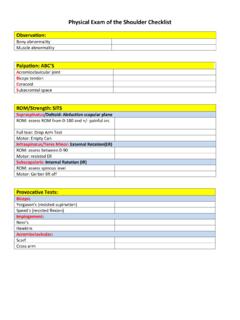
3 Strength, neuro-vascularassessment. Specific provocative maneuvers If acute injury& pain difficultto assessas patient protects limiting movement, examination Examineunaffected side first(gain confidence, develop sense of theirnormal)Knee Anatomy:Hinge Type Joint Logical ExamImages courtesy of Dr. Ted Parks,Western OrthopaedicsPutting It All TogetherImages courtesy of Dr. Ted Parks,Western OrthopaedicsObservation Obvious pain or gait abnormality? Redness or other discoloration? Scars past surgery? Swelling fluid in the joint (aka effusion)?
4 Atrophic muscles ( from chronic disuse)? Alignment: Bowing of legs (inward =s Valgus, outward =s Varus)?Varus Deformity (bowing outward)SurgicalScarsObvious right knee effusionValgus Deformity (bowing outward)Palpation: Patellar Mechanism Fully expose take off pants, use gown or shorts! Palpate note any warmtharound knee quadriceps& hamstringmuscle groups patella(knee cap) quadriceps and patellar tendons anterior tibial tuberosity (insertion patellar tendon)HamstringMusclesPatellar PalpationPatellofemoral Anterior knee pain secondary to patella articulation w/femurTo Test: Slightly flex knee.
5 Patellar Apprehension Test: Move patella side to side if too much laxity patient will fear subluxation Palpate patella facets: May elicit pain if Chondromalacia. Patella Grind (aka Quad Apprehension Test): Examiner pushes down on patella while patient contracts quadriceps forces patella onto femur, eliciting pain behind knee cap in Patellofemoral syndromeCourtesy Orthopedic Specialists of of Motion (ROM) Active then passive (you move the joint) Hand on patella w/extension & flexion osteoarthritis, may feel grinding sensation (crepitus)Normal range of motion:Full Flexion: 1400 Full Extension.
6 00 Joint Line PalpationJoint Line Tenderness medial or lateral meniscal injury & Osteoarthritis (OA) Slightly flex knee. Find joint space along lateral & medial margins. Joint line perpendicular to long axis tibia. Palpate along medial, then lateral margins. Pain suggest underlying meniscus damage or OALateralMedialMenisci Normal Function and Anatomy Medial & lateral menisci on top of tibia cushioned articulating surface between femur & tibia Provides stability, distributes force & protects underlying articular cartilage (covers bone, allows smooth movement) Menisci damaged by trauma or degenerative changes w/age.
7 Symptoms if torn piece interrupts normal smooth movement of joint pain, instability ("giving out"), locking &/or swellingCourtesy U of of the MenisciWithout MeniscusWith MeniscusFemurTibiaFibulaCourtesy Orthopedic Specialists of CollateralLigamentLateral CollateralLigamentAdditional Tests For Meniscal Injury McMurray s Test Medial MeniscusMcMurray s manipulates knee torn meniscus pinched pain & clickMedial meniscus: Left hand w/middle, index, & ring fingers on medial joint line. Grasp heel w/right hand, fully flex knee. Turn ankle foot pointed outward (everted), knee pointed outward.
8 Holding foot in everted position, extend & flex knee. If medial meniscal injury, feel "click" w/hand on knee w/extension. May also elicit pain. Simulated McMurray s Note pressure placed on medial meniscusMcMurray s Test Lateral Meniscus Return knee to fully flexed position, turn foot inwards (inverted). Direct knee so pointed inward. Hand on knee, fingers along joint lines Extend and flex knee. If lateral meniscal injury feel "click" w/fingers on joint line; May also elicit : McMurray s Test for medial and lateral meniscus injuries are performed togetherLigaments Normal Anatomy and Function 4 bands tissue, connecting femur tibia provide stability MCL, LCL, ACL, PCL Ligamentous injury.
9 Requires significant force can be non-contact acute pain, swelling & often hear a "pop" (sound of ligament tearing) longer term instability (give-way)Courtesy U of CollateralLigamentLateral CollateralLigamentFemurTibiaFibulaSpecif ics of Testing Medial Collateral ligament (MCL) Flex knee ~ 300 Left hand on lateral aspect knee. Right hand on ankle or calf. Push inward w/left hand (Valgus force). If MCL torn, joint "opens up" along medial aspect. May also elicit pain w/direct palpation over ligamentCompare w/non-affected side normal laxity varies from patient to patientDirection of ForceDirection of ForceImages courtesy of Dr.
10 Ted Parks,Western OrthopaedicsLateral Collateral ligament (LCL) Flex knee ~ 300 Right hand medial aspect knee. Right hand on ankle or calf. Push steadily w/left hand (Varus force) If LCL torn, joint will "open up" on lateral aspect. May elicit pain on direct palpation of injured ligamentDirection of ForceDirection of ForceImages courtesy of Dr. Ted Parks,Western OrthopaedicsAnterior cruciate ligament (ACL) Lachman s Test Grasp femur w/left hand, tibia w/right. Flex knee slightly. Pull up sharply (towards belly button) w/right hand, stabilizing femur w/left.