Example: barber
Musculoskeletal Ultrasound Technical Guidelines IV
band. Shifting the probe posteriorly, the anterior portion of the gluteus maximus can be seen covering the posterior part of the tendon of the gluteus medius. Coronal planes demonstrates the fascia lata which appears as a superficial hyperechoic band that, from cranial to caudal, overlies the gluteus medius muscle,
Tags:
Information
Domain:
Source:
Link to this page:
Documents from same domain

Musculoskeletal Ultrasound Technical Guidelines I.Shoulder
www.essr.orgDynamic assessment of subacromial (antero-superior) impingement can be attempted by placing the probe in the coronal plane with its medial margin at the lateral margin of the acromion. The patient abducts his arm while in internal rotation. With this manoeuvre, the supraspinatus and the bursa can be seen passing deep to the coracoacromial arch ...

Musculoskeletal Ultrasound Technical Guidelines V
www.essr.orgfield-of-view of the US image. Much gel may help to avoid excessive pressure on the bursa with the probe. 4 With patient’s positioning described at point-1, examine the patellar tendon from its cranial origin down to its distal insertion using long- and short-axis planes. Because the lower pole of the patella has a V-shaped appearance,
Musculoskeletal Ultrasound Technical Guidelines III
www.essr.orgMusculoskeletal Ultrasound Technical Guidelines III.Wrist Ian Beggs, UK Stefano Bianchi, Switzerland Angel Bueno, Spain Michel Cohen, France Michel Court-Payen, Denmark ... extensor tendons can be performed by placing the hand on a gel tube with the fingers hanging outside its edge to allow easy fingers movements. 1 Wrist

Musculoskeletal Ultrasound Technical Guidelines VI
www.essr.orgExamine the flexor digitorum longus tendon down to reach the sustentaculum tali. Look at the flexor retinaculum, the posterior tibial vessels and the tibial nerve with its divisional branches (medial and lateral plantar nerves). Compression may help to assess whether the veins are patent. Legend: AbdH, abductor hallucis muscle; curved arrow,
Related documents

ROSE K2 Keratoconus Fitting Tips - Menicon
cdn.menicon.nlEDGE BAND TOO NARROW ... so the change in edge lift (which alters the sagittal height) does not affect the central fit! With ROSE K2 lenses, 85% of all lenses dispensed use either the standard edge, standard flat (increased) or standard steep (decreased) edge lift to achieve the desired peripheral fit. However, other edge lift values can be ...
BASIC APPROACH TO EVALUATING A HEAD CT - Brigham and …
www.brighamandwomens.orgBlue arrow – band of streak artifact limits evaluation of the pons . CT Neuroimaging The head is routinely scanned using sequential imaging in the axial plane with ... sagittal sinus . Anatomy Yellow – centrum semiovale are supraventricular white matter tracts running to …
Oral Surgery: Orthodontic Related Procedures
www.uhcprovider.comThis is the placement of an orthodontic bracket, band or other device and attached with a chain, on an unerupted tooth, after surgical exposure, to aid in its eruption. This procedure is done following the surgical access of an unerupted tooth. ... A cephalometric sagittal occlusion analysis merged with mandibular incisor

Sagittal Band Injury - Emory University School of Medicine
med.emory.eduSagittal Band Rupture •Also known as traumatic extensor tendon dislocation and boxers knuckle •Mechanism of injury –Most commonly occurs in flexed position with when a knuckle hits a sharp surface (i.e. tooth) resulting in an oblique laceration (central laceration may lead to isolated injury to the extensor tendon) •Location
Musculoskeletal Ultrasound Technical Guidelines I.Shoulder
www.essr.orgshifted-down) on sagittal planes. 8 6 Shoulder Look at the infraspinatus and teres minor muscles as individual structures filling the in-fraspinous fossa deep to the deltoid. After scanning these muscles, sweep the transduc-er toward the greater tuberosity on sagittal planes. The two tendons can be appreciated
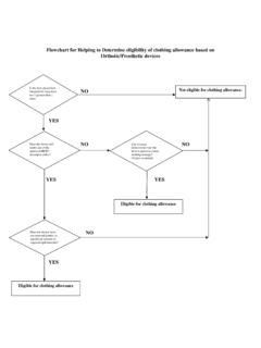
NO Not eligible for clothing allowance. YES YES YES
www.prosthetics.va.govL0468 - TLSO, sagittal-coronal control, rigid posterior frame and flexible soft anterior apron with straps, closures and padding, extends from sacrococcygeal junction over scapulae, lateral strength provided by pelvic, thoracic, and lateral frame pieces, restricts gross trunk motion in sagittal, and coronal planes, produces
GLOSSARY of MEDICAL and ANATOMICAL TERMS
medsci.indiana.eduarachnoid granulations protrusions of arachnoid into superior sagittal sinus whereby cerebrospinal fluid can pass into the blood, cf. Pacchionian bodies. arbor vitae L. arbor = tree + vitae = of life; cedar tree; the white matter seen in a median section of the cerebellum. arbor vitae uteri cf. palmate folds (of uterine cervical canal).
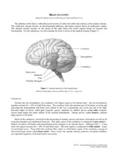
BRAIN ANATOMY - WOU
people.wou.eduBI 335 – Advanced Human Anatomy and Physiology Western Oregon University Figure 4: Mid-sagittal section of brain showing diencephalon (includes corpus callosum, fornix, and anterior commissure) Marieb & Hoehn (Human Anatomy and Physiology, 9th ed.) – Figure 12.10 Exercise 2: Utilize the model of the human brain to locate the following structures / landmarks for the