Transcription of Non-Invasive Fourier Transform Infrared Microspectroscopy ...
1 Modern Research and Educational Topics in Microscopy. FORMATEX 2007 A. M ndez-Vilas and J. D az (Eds.). _____. Non-Invasive Fourier Transform Infrared Microspectroscopy and imaging Techniques: Basic Principles and Applications P. Garidel*1, and M. Boese2. 1. Institute of Physical Chemistry, Faculty of Chemistry, Martin-Luther-University Halle/Wittenberg, Muehlpforte 1, D-06108 Halle/Saale, Germany 2. Bruker Optics GmbH, Rudolf-Plank-Str. 27, D-76275 Ettlingen, Germany The current paper describes rather new and straightforward Non-Invasive mid Infrared spectroscopic techniques for the position sensitive identification of sample components. These techniques include mid Infrared (MIR) Microspectroscopy , mapping as well as imaging approaches in transmission, reflection-absorption, or attenuated total reflection mode. The basic principles of the different Infrared Microspectroscopy and imaging techniques are presented, with a critical evaluation of the technique compared to currently utilized other imaging techniques.
2 The presented examples are focussed on applications in life science, exploration of the heterogeneity of a single sample, identification and biochemical characterisation of biological samples, ex-vivo monitoring the distribution of drugs in tissues. Additional applications are briefly discussed to illustrate the potentials and limitations of the presented techniques for medical diagnostics. Keywords Infrared Microspectroscopy ; mapping; imaging ; Non-Invasive ; tissue; FTIR. 1. Introduction The coupling of an Infrared spectrometer to an optical microscope (Figs. 1 and 2) offers the unique opportunity for studying biological samples with a spatial resolution limited by the near diffraction limit of Infrared light (3-10 m). The development efforts of the last years have enlarged the potential applications of Infrared microspectroscopic analysis, since measurements can be performed in transmission (Figs.)
3 3 - 5), reflection-absorption, diffuse reflection, grazing incidence reflection and attenuated total reflection (ATR) mode, which can be associated to different micro and macro sampling accessories (Fig. 6). Additionally, the microspectroscopic imaging instruments include features enabling the conduction of polarisation experiments, experiments using differential interference contrast, or allowing Normaski measurements [11, 12]. The examination of the spectral data can be performed as univariate analysis, however the use of multivariate analysis allows the extraction of much more information [1 4]. Therefore, large efforts are also undertaken to develop adequate chemometry tools for data evaluation [5 10]. The derived information from Infrared Microspectroscopy is mostly presented as a false colour image, which resembles histological staining samples. The purpose of this article is to provide the basic knowledge required to conduct Infrared Microspectroscopy mapping and imaging experiments.
4 Instrumentation setups as well as data analysis are briefly discussed. For introductions to microscopy or vibrational spectroscopy it is referred to educational textbooks [11, 12]. Various examples (unpublished) in the field of life science are presented to demonstrate the applicability of different Microspectroscopy mapping and imaging experimental modes. *. Corresponding author: e-mail: 132. Modern Research and Educational Topics in Microscopy. A. M ndez-Vilas and J. D az (Eds.) FORMATEX 2007. _____. 2. Basic Principles Infrared Microspectroscopy mapping and imaging techniques are Non-Invasive approaches in the sense that no extrinsic staining is required to generate the image contrast, as it is applied in fluorescence imaging spectroscopy. In fluorescence microscopy imaging [13] only the used fluorescence dyes are visualised in the sample, and basically no information with regard to the biochemical composition of the sample is derived.
5 Using vibrational spectroscopy approaches intrinsic spectroscopic properties of the sample itself are monitored. Raman Microspectroscopy [14-16], a spectroscopy technique that is complementary to Infrared spectroscopy, has evolved fast in the last years, since Raman technology has largely benefited from the advancements in laser as well as array detector developments. However, using the Raman approach it has to be considered that possible sample damage may occur due to the use of strong laser intensities. This is especially a problem for the analysis of biological samples. Additionally, limited applications may result from interferences with fluorescence of the sample and the signal arising from Raman scattering cross sections is typically six orders of magnitude smaller compared to Infrared absorptivities [14]. However, the correct choice of the used spectroscopic technique depends strongly on the aim of the study (analysed sample, sensitivity of the sample to the procedure, required lateral resolution etc.)
6 Single element detector Focal plane array (FPA). detector Measuring modes (transmission, reflection, ATR). Motorized stage (x-y). FTIR spectrometer microscope Fig. 1 Fourier Transform mid Infrared microspectroscopic instrumentation (Hyperion 3000). FTIR: Fourier Transform Infrared , ATR: attenuated total reflection. Infrared mapping approach The first investigations, in the early 1990's, employed FTIR ( Fourier Transform Infrared ). Microspectroscopy instruments equipped with a single Infrared point detector, enabling just to analyse a single section, single spot, defined by the aperture of the microscope [17, 18] (Fig. 2a). In order to get a high lateral resolution the minimal aperture dimension is set to approximately 10 m x 10 m. With this set-up it is possible to investigate spectroscopically single cells, which allows the classification of normal versus malignant cells in medical diagnostic analysis [10, 32, 34, 43].
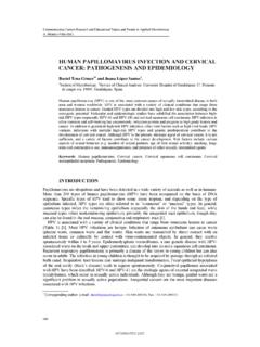
7 133. Modern Research and Educational Topics in Microscopy. FORMATEX 2007 A. M ndez-Vilas and J. D az (Eds.). _____. A B C. Spot #1. Spot #2. sample Analysed area Single pixel IR image FPA element D IR spectrum E. high Intensity Intensity low 4000 3500 3000 2500 2000 1500 1000. -1. Wavenumber / cm Fig. 2 Principle of Infrared microspectroscopic mapping and imaging . (A) Optical micrograph of the sample being analysed using the mapping approach, (B) schematic representation of a focal plane array (FPA) detector made of a large number of pixel elements for the investigation of the sample area of interest, (C) Infrared image of the analysed sample representing the lateral distribution of a chemical entity, (D) Infrared spectrum obtained from a single pixel element of the FPA detector, (E) Infrared image colour code: red corresponds to a high amount (intensity), blue corresponds to a low amount (intensity) of the imaged chemical entity.
8 The mapping approach was the first technique developed for the lateral spectroscopic characterisation of larger samples. The spatially resolved spectral information is obtained by scanning the sample spot by spot across the surface using a stationary detector and collecting at each spot a complete Infrared spectrum (Fig. 2A). The sample is moved by an x-y-computer controlled stage, in order to position the next spot under the microscope. The spot registered at each step across the sample is defined by the aperture of the microscope, which may ranges between 10 m x 10 m up to 200 m x 200 m. The mapping procedure permits the acquisition of high quality Infrared spectra with spatial information, which are used to map the distribution of chemical entities in the sample. The different spots are recorded sequentially, and the image is reconstituted by matching the spectral information of each single spot to construct a larger image.
9 The drawback of this procedure is that the investigation of larger sample areas, using a reasonable lateral resolution (between 10-40 m), is extremely time consuming (taking up to days for larger samples in the mm range analysed at a lateral resolution of 10-40 m) [19]. 134. Modern Research and Educational Topics in Microscopy. A. M ndez-Vilas and J. D az (Eds.) FORMATEX 2007. _____. The obtained spatial resolution for FTIR microscopy is limited by the diffraction limit of the Infrared light. The diffraction limited spatial resolution (d) depends on the wavelength of the used light ( ), the refractive index of the medium in which the measurement is performed (n') and the angle ( ) defining the most extreme ray exciting the optical system with respect to the central axis and thus is given by: d = ( ) / (n' sin ) [11]. For most Infrared microscope objectives the numerical aperture NA (NA = n' sin ) ranges between and Thus the diffraction limited spatial resolution in the Infrared finger print region (4000 1000.)
10 Cm-1) is 3-10 m. Lasch and Naumann [37] have recently pointed out that the spatial resolution is one of the most critical measurement parameters in FTIR Microspectroscopy . Due to the distinct levels of morphologic heterogeneity in cells and tissues the spatial resolution in a given IR imaging setup strongly affects the character of the Infrared spectral pattern obtained from the biomedical sample. These authors have presented a computational procedure, three-dimensional Fourier self deconvolution, which is suitable to improve the spatial resolution in Infrared imaging (for more details see [37]). Infrared imaging approach The breakthrough using Infrared Microspectroscopy was the accessibility of array detectors [20-25]. The use of a focal plane array (FPA) detector instead of using a single element detector reduced the experimental time drastically for the investigation of a larger sample area (Fig.