Transcription of Optical and Structural Properties of Zinc Oxide Nanoparticles
1 International Journal of Advanced Research in Physical Science (IJARPS). Volume 2, Issue 1, January 2015, PP 14-18. ISSN 2349-7874 (Print) & ISSN 2349-7882 (Online). Optical and Structural Properties of zinc Oxide Nanoparticles Swati S. Kulkarni Mahavidyalaya Beed Department of Physics Beed City, (MS) INDIA. Mahendra D. Shirsat Intelligent Material Research Laboratory Department of Physics Dr. Babasaheb Ambedkar Marathwada University Aurangabad, Aurangabad (MS) INDIA. Abstract: In the recent era zinc Oxide Nanoparticles attracted the researchers due to their unique Properties and applications in optoelectronic devices. zinc Oxide Nanoparticles are the semiconductor materials having band gap energy eV and very large excitation binding energy (60meV) at room temperature. Also it is non-toxic, environmental friendly and transparent to visible range of spectrum. In the present investigation zinc Oxide Nanoparticles were synthesized by sol gel method using zinc acetate and sodium hydroxide as precursors and de ionized (DI) water as solvent.
2 The structure and morphology of prepared zinc Oxide Nanoparticles was studied using X-ray diffraction (XRD) spectroscopy and Scanning Electron Microscopy (SEM). UV visible spectrum shows the transparency of Nanoparticles over entire visible range. The FTIR analysis confirms the formation of zinc Oxide Nanoparticles . Keywords: zinc Oxide Nanoparticles , sol-gel, XRD, SEM, UV visible spectra. 1. INTRODUCTION. zinc Oxide (ZnO) Nanoparticles have become famous among researchers due to its use in various applications like gas sensors [1], chemical sensors [2-3], biosensors [4-5], superconductors [6], photo catalyst [7], optoelectronic devices[8-10],cosmetics[11],etc. ZnO is a wide band gap semiconductor having high Optical transparency and Luminescence in visible and near ultraviolet range of spectrum. Therefore, it is usually used in light emitting diodes and solar cells. ZnOnano particles are having high exciton binding energy nearly 60 meV. This means the excitonic transition in case of ZnO Nanoparticles is possible at room temperature also [12-14].
3 Moreover zinc Oxide is environmental friendly and ease to synthesize [15]. Many techniques are being used to synthesize ZnO Nanoparticles Viz. precipitation method, spray pyrolysis method, micro emulsion method, hydrothermal method and Sol gel method [16 20]. In the present investigation the sol-gel method for synthesis of ZnO Nanoparticles was chosen as it is simplest method, consumes less power and can be carried out in robust atmosphere. The Structural Properties of as prepared ZnO Nanoparticles was studied by using X Ray Diffraction(XRD) and the morphology of ZnO Nanoparticles was examined under Scanning Electron Microscope (SEM). The transparency and absorption of synthesized ZnO Nanoparticles was studied using UV visible spectrophotometer. The formation of ZnO Nanoparticles was confirmed from FTIR analysis. 2. METHODS. All the reagents utilized were of analytical grade purchased from Rankem India Pvt. Ltd. and used as it is without further purification.
4 Gm of zinc acetate dehydrate was dissolved in 100 ml de- ionized water by stirring for half an hour. 4 gm sodium hydroxide pellets were dissolved in 100. ml de-ionized water by stirring 15 minutes. This solution was added drop by drop to above prepared zinc acetate solution to achieve the pH nearly 12. The pH test was carried out with the ARC Page | 14. Swati S. Kulkarni & Mahendra D. Shirsat help of pH tester. The solution was further stirred for one hour and precipitate was obtained. Collected precipitate was washed many times by de-ionized water, filtered and dried overnight in the hot air oven at 60 degree Celsius. The fine powder was made by grinding the dried precipitate before using for characterization. The morphology of the ZnO Nanoparticles was examined with the help of Hitachi Model S-300H. SEM machine. The structure and size of the ZnO Nanoparticles was studied with the help of Bruker AXS D8 Advance X Ray Diffractometer using Cu-K radiations having wavelength The UV visible spectrum of the sample was obtained using a Shimatzo's UV visible spectrophotometer-1800.
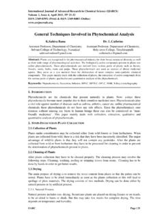
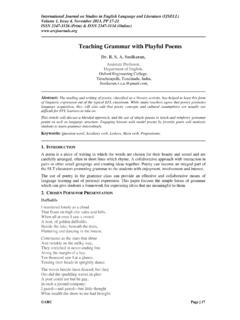
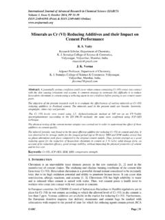
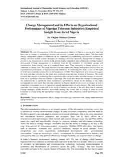
5 3. RESULTS AND DISCUSSION. Structural Properties The X Ray Diffraction spectrum of synthesized ZnO Nanoparticles shown in figure 1 confirms the hexagonal wurtzite structure according to JCPDF data (card number PDF#800075 ICDSD#. 067849). Wurtzite structure of ZnO Nanoparticles is belonging to space group (C6V=P63mc) and having unit cell parameters a = b = and c = . The average size of the Nanoparticles has been calculated for the 101 peak using the Debye-Scherrer formula, Where, is the x-ray wavelength ( nm), is the Bragg diffraction angle, and is the full width at half maximum. The full width at half maximum was measured using Gaussian curve for the highest Peak 101. The average size of Nanoparticles was found to be equal to nm. Fig1. XRD pattern of ZnO Nanoparticles Morphological Study The SEM image of the sample is shown in fig 2(a) and 2(b) reveals that the particles are spherical and having granular nature. In the higher resolution SEM image the agglomeration of particles is observed.
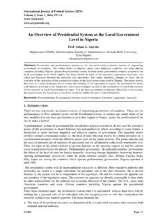
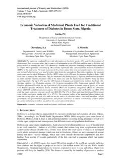
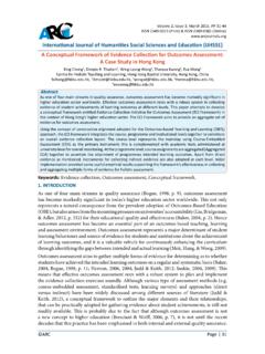
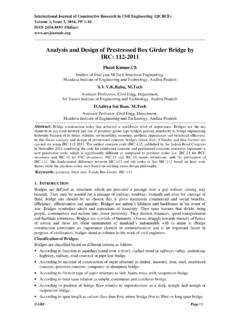
6 Aging may have caused the agglomeration. Fig2 (a). SEM image of ZnO Nanoparticles International Journal of Advanced Research in Physical Science (IJARPS) Page | 15. Optical and Structural Properties of zinc Oxide Nanoparticles Fig2 (b). SEM image of ZnO Nanoparticles with higher resolution Optical Study The UV visible spectrum of the sample is as shown in fig 3. The sample absorbs the radiations in the UV range up to and almost all the visible spectrum radiations are transmitted by the ZnO Nanoparticles . The band gap of the ZnO Nanoparticles was calculated by extrapolating the curve drawn between (h ) and ( h ) 2(shown in fig 4). Where is the frequency and is the Optical absorption coefficient. The band gap energy obtained by extrapolating curve is found to be approximately Fig3. UV visible absorption spectra Fig4. Extrapolation curve for band gap determination of as synthesized ZnO Nanoparticles FTIR curve for ZnO Nanoparticles is shown in fig 5.
7 The secondary vibrations of Zn-O bond are found to be lying at per cm. The peaks lying from 1500 to 1700, 2300 to 2700 and at 3000. per cm are representing the functional groups corresponding to C-O symmetric and anti- International Journal of Advanced Research in Physical Science (IJARPS) Page | 16. Swati S. Kulkarni & Mahendra D. Shirsat symmetric stretching mode, C-H stretching mode and O-H stretching mode respectively. Presence of O-H group represents the presence of water molecules on the surface of ZnO Nanoparticles . Fig5. FTIR spectrum of ZnO Nanoparticles 4. CONCLUSION. ZnO Nanoparticles were synthesized by simple sol-gel method. The secondary bond vibration at per cm in FTIR spectrum confirms the production of ZnO Nanoparticles . The XRD. examination verified the production of wurtzite shape Nanoparticles . SEM image confirms the formation of spherical granular ZnO Nanoparticles . The absorption peak is found to be at nm. The energy band gap of as synthesized Nanoparticles was obtained to be equal to eV.
8 REFERENCES. [1] S K Gupta, Aditee Joshi and Manmeet Kaur, Development of gas sensors using ZnO. nanostructures, J. Chem. Sci. 122,57 (2010). [2] Zhiyong Fan and Jia G. Lu, Chemical Sensing with ZnO Nanowires, IEEE 834, (2005). [3] Zhiyong Fan and Jia G. Lu, Chemical Sensing With ZnO Nanowire Field-Effect Transistor, IEEE Transactions On Nanotechnology 5, 303 (2006). [4] Zhiwei Zhao, Wei Lei, Xiaobing Zhang, Baoping Wang and HelongJian, ZnO-Based Amperometric Enzyme Biosensors, Sensors 10, 1216(2010). [5] XU ChunXiang, YANG Chi, GU BaoXiang& FANG ShengJiang, Nanostructured ZnO for biosensing applications, Chin Sci. Bull. 58, 2563 (2013). [6] Parmanand Sharma, Amita Gupta, Rao, Frank J. Owens, Renu Sharma, Rajeev Ahuja,J. M. Osorio Guillen, B rjeJohansson and G. A. Gehring, Ferromagnetism above room temperature in bulk and transparent thin films of Mn-doped ZnO,nature materials, 2, 673. (2003). [7] AbdulazizBagabas, Alshammari, Mohmed FA Aboud and HendricKosslick, Room temperature synthesis of zinc Oxide Nanoparticles in different media and their application in cyanide photo-degradation, nanoscale Research letters, 8, 516 (2013).
9 [8] Shuyan Shao, KaiboZheng, KarelZidek, PavelChabera, TonuPullerits and Fengling Zhang, Optimizing ZnO nanoparticle surface for bulk heterojunction hybrid solar cells, Solar Energy Materials and Solar Cells, 118, 43 (2013). [9] S. VenkataprasadBhat, A. Govindaraj, C. N. R. Rao, Hybrid solar cell based on P3HT ZnO. nanoparticle blend in the inverted device configuration, Solar Energy Materials & Solar Cells, 95, 2318 (2011). [10] Yunfei Zhou, Michael Eck and Michael Kr ger, Organic-Inorganic Hybrid Solar Cells:State of the Art, Challenges and Perspectives, Solar Cells New Aspects and Solutions, 95. (2011). [11] Mohammad Vaseem, Ahmad Umar, Yoon-Bong Hahn, Metal Oxide Nanostructures and Their Applications, American Scientific Publishers (2010) ch4,Pp 1 36. International Journal of Advanced Research in Physical Science (IJARPS) Page | 17. Optical and Structural Properties of zinc Oxide Nanoparticles [12] Soosen Samuel M, Lekshmi Bose and George KC, Optical Properties of ZnO Nanoparticles , SB Academic Review, 16(1 & 2), 57 (2009).
10 [13] Peng W Q, Qu S C, Cong G W and Wang Z G, Structure and visible luminescence of ZnO. Nanoparticles , Mater. Sci. Semic. Proc., 9,156 (2006). [14] Yu Q, Fu W, Yu C, Yang H, Wei R, Sui Y, Lui Y, Lui Z, Li M, Wang G, Shao C, Lui Y and Zou G, Structural Electrical and Optical Properties of Yttrium doped ZnO Nanoparticles , J. Phys. D: Appl. Phy.,40,5592 (2007). [15] Wu, Tok, Boey, Zeng, Zhang, Surface modification of ZnOnanocrystals, Applied Surface Science, 253, 5473 (2007). [16] Chen S, Kumar RV, Gedanken A, Zaban A, Sonochemical Synthesis of crystalline nanoporous zinc Oxide Spheres and their application in Dye Sensitized Solar Cells, Isr. J. Chem., 41,51 (2001). [17] Ghaffarian, Hamid Reza; Saiedi, Mahboobeh; Sayyadnejad, Mohammad Ali, Synthesis of ZnO Nanoparticles by Spray Pyrolysis Method, Iran. J. Chem. Chem. Eng.,30, 1 (2011). [18] Kelly Pemartin, conxitasolans, German Vidal-Lopez and Margarita Sanchez-Dominguez, synthesis of ZnO and ZnO2 Nanoparticles by the oil-in-water micro emulsion reaction method, , 41,1032 (2012).