Transcription of Oxygen Saturation: A Guide to Laboratory Assessment
1 10 CLINICAL Laboratory NEWS FEBRUARY 2006 CLN SLAB 2006:SUPPORTING CLINICALDECISIONSSERIESThe Clinical Laboratory Standards In-stitute (CLSI) defines O2sat as an estimated value based on a calculation, whereas SaO2and SpO2 refer to the arterial saturation measured spectrophotometrically. (To avoid further confusion, I will use only O2sat and SO2 for the remainder of this article.) CO-oximetry, a more complex and reliable method, measures the concentration of he-moglobin derivatives in the blood, the results of which are then used to calculate various parameters such as hemoglobin derivative fractions ( , FO2Hb), total hemoglobin, and Oxygen most patients, the results O2sat, SO2, and FO2Hb from these three methods are virtually identical. But in cases of dyshemo-globinemia, some methods can yield mis-leading results, so health care professionals need to understand the differences between these methods and the limitations of each.
2 This article will review the basics of Oxygen -ation of hemoglobin and will provide guid-ance for laboratorians on how to accurately assess oxygenation saturation Oxygen saturation PrimerHemoglobin, the O2 transport protein in blood, is comprised of four subunits: two subunits and two non- subunits, for example , , or . Each subunit contains a porphyrin heme iron (Fe) moiety and seven helices. The heme Fe exists in the Fe(II) or Fe(III) oxidation state, but only the Fe(II) state is capable of binding O2. Most clini-cally important dyshemoglobinemia gene mutations, such as the thalessemias, reduce the quantity of - or -chain synthesis in the hemoglobin subunit or lower the solubility of hemoglobin as occurs with HbS (sickle cell hemoglobin) or HbC (hemoglobin C), for example but gene mutations rarely al-ter the O2 affinity of can be divided into two classes: normal hemoglobin that is capable of binding O2, and dyshemoglobins he-moglobin derivatives that are incapable of binding O2.
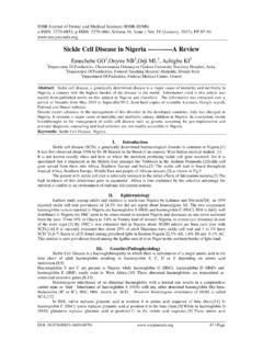
3 The normal hemoglobins in-clude oxyhemoglobin (O2Hb) and deoxy-hemoglobin (HHb), while the dyshemoglo-bins include carboxyhemoglobin (COHb), methemoglobin (MetHb), and sulfhemo-globin (SHb). COHb forms when a person is exposed to CO fumes and CO replaces O2 in hemoglobin, which can result in death. SHb forms through a reaction of sulfa-contain-ing compounds with the heme moiety; cases of sulfhemoglobinemia are rare and usually result from extensive use of sulfa-containing drugs. MetHb represents the oxidized, deoxy form (Fe(III)-Hb) of hemoglobin to which O2 cannot bind. The likely etiologies of met-hemoglobinemia include exposure to highly oxidizing drugs or the presence of genetic hemoglobin variants such as understand how hemoglobin carries and releases Oxygen , the Oxygen dissociation curve (ODC) serves as an important tool (Figure 1). In the lungs, where partial pres-sures of O2 are high, O2 binds to hemoglo-bin to form O2Hb.
4 Erythrocytes carrying O2Hb then circulate in the blood and release O2 in response to decreased partial pressures of O2 in the tissues. The cooperativity of O2binding an allosteric phenomenon where-by binding of Oxygen by one hemoglobin subunit enhances the ability of the remain-ing subunits to bind Oxygen produces the sigmoidal shape of the curve. As O2 binds to the second and third subunits of hemoglo-bin, binding increases incrementally so that the four subunits of hemoglobin all become fully saturated at the normal O2 tension in Oxygen saturation A Guide to Laboratory AssessmentBY SHANNON HAYMOND, PHDH uman life depends on the Oxygen transport by hemoglobin. In healthy patients, the major-ity of molecular Oxygen (O2) is bound to hemoglobin and only a small fraction is dissolved in blood. But in patients with respiratory problems or certain metabolic and genetic disorders, the fraction of oxygenated hemoglobin can fall to dangerously low values.
5 Therefore, labora-tory Assessment of Oxygen saturation (SO2) the percentage of hemoglobin saturated with Oxygen provides an important indicator of a patient s cardio-respiratory status and is frequently used in the emergency department, during general and regional anesthesia, and in intensive care the measured parameters are quite different for each, the three major analytical methods for mea-suring Oxygen saturation arterial blood gas analyzers, pulse oximetry, and CO-oximetry are frequently used interchangeably by health care workers. Arterial blood gas analyzers calculate estimated Oxygen saturation (O2sat) in a blood sample based on empirical equations using pH and PO2 values, while pulse oximeters moni-tor arterial blood Oxygen saturation , commonly referred to as SpO2 or SaO2, noninvasively by passing selected wavelengths of light through an area of the body, such as a finger.
6 Both are measures of Oxygen saturation . CLINICAL Laboratory NEWS FEBRUARY 2006 11lung alveoli. The same process works in re-verse in the tissues; once fully loaded hemo-globin releases one O2 molecule, it releases the next more ODC also explains what happens to patients when Oxygen cannot bind hemoglo-bin. For example, in the presence of CO or when the heme Fe is oxidized to the Fe(III) state, O2 cannot bind one of the hemoglo-bin subunits. These conditions decrease O2 capacity in addition to inhibiting O2 trans-port by blood. In addition, CO and oxidized heme Fe alter hemoglobin s conformation in a way that decreases the O2 affinity for the remaining heme Fe groups, shifting the ODC to the right. The net biological effect is decreased O2 delivery to tissues. Tem-perature, pH, and 2,3-diphosphoglycerate (2,3-DPG) concentration also affect the O2 affinity of hemoglobin, shifting the ODC as shown in Figure important aspect of the ODC is the P50, the partial pressure of Oxygen in the blood at which the hemoglobin is 50% satu-rated.
7 The value for P50 typically about mm Hg for a healthy person is used as an indicator of the affinity of hemoglobin for O2. In the presence of disease or other conditions that change the hemoglobin s affinity for O2, the curve shifts to the right or to the left and the P50 changes accord-ingly. An increased P50 indicates a rightward shift of the standard curve and means that a higher partial pressure is necessary to main-tain 50% Oxygen saturation . Increases in temperature and 2,3-DPG concentration, or decreases in pH, can shift the ODC to the right, increasing the P50 and indicating de-creased O2 of Oxygenation StatusCLSI has defined the three key terms used to describe oxygenation status: Oxygen sat-uration (SO2), fractional oxyhemoglobin (FO2Hb), and estimated Oxygen saturation (O2sat). The Institute recommends the use of the term Oxygen saturation to indi-cate the amount of hemoglobin capable of transporting O2 and fractional O2Hb to represent the fraction of hemoglobin that is oxygenated.
8 The terms fractional satura-tion and functional saturation refer to the FO2Hb and SO2, respectively. Oxygen saturation . The following empiri-cal equations are used to determine Oxygen saturation , SO2, from measured hemoglo-bin concentrations:Total hemoglobin concentrationctHb = cO2Hb + cHHb + cMetHb + cCOHb + cSHbHemoglobin Oxygen saturation SO2 = cO2Hb cO2Hb + cHHbPulse oximetry and CO-oximetry report SO2, but with these methods, the values are often referred to as SaO2 or SpO2. Regardless of the method used, SO2 is a measure of the fraction of oxygenated hemoglobin in rela-tion to the amount of hemoglobin capable of transporting O2. The normal range for SO2, expressed as a percentage, is typically 94% 98%. Blood gas analyzers report an estimated saturation , O2sat, which is based on measurement of pH, PO2, and hemoglo-bin values and utilization of empirical oxyhemoglobin.
9 Only instru-ments with a multi-wavelength spectro-photometer, such as CO-oximeters or some modern blood gas analyzers, are capable of measuring FO2Hb. The value, as calculated from the formula below, represents the frac-tion of oxyhemoglobin in relation to the to-tal hemoglobin present, including the non- Oxygen -binding oxyhemoglobinFO2Hb = cO2Hb ctHbFO2Hb is usually expressed as a percent-age and typically ranges from 90% 95% in healthy individuals. Reporting FO2Hb alone is of limited value because the dyshemoglo-bin fractions also play an important role in the analysis of Oxygen carrying capacity. The fraction of any of the hemoglobin deriva-tives may be calculated in the same manner as that used for demonstrated by the above equations, SO2 and FO2Hb are not equivalent terms. In healthy individuals, the values obtained for SO2 and FO2Hb are approximately equal, due to the absence of dyshemoglobins.
10 For patients with dyshemoglobinemias, how-ever, FO2Hb will be considerably lower than SO2. Although the SO2 typically remains within the normal range in the presence of elevated COHb or MetHb, the O2 capacity may be severely decreased, leading to fatal Assessment Of Oxygenation StatusHemoglobin molecules are easily measured by spectrophotometric methods. In addi-tion, the altered molecular structure of the heme moiety in the various hemoglobin derivatives gives rise to unique absorption spectra, making it possible to determine the concentrations of each derivative present in blood. (Figure 2)Pulse oximetry. Pulse oximeters assess arte-rial Oxygen saturation (SO2) by measuring light transmission through a well-perfused area of the body such as a finger or an ear lobe. Light sources, typically light-emit-ting diodes, shine two wavelengths of light through the tissue visible red (R) light (600 to 750 nm) and infrared (IR) light (850 to 1000 nm).