Transcription of Pacemaker Learning Package - Agency for Clinical Innovation
1 Pacemaker Learning Package Paula Nekic CNE. Liverpool Hospital ICU. January 2016. Liverpool Hospital Intensive Care: Learning packages Intensive Care Unit Pacemaker Learning Package CONTENTS. Indications for pacing 3. Anatomy 4. Definitions 9. Types of pacing 13. How does a Pacemaker work? 20. Modes of pacing 24. Sensitivity and output 32. Thresholds 35. Management 37. Precautions 40. Complications / troubleshooting 41. Learning activities 45. Reference List 46. LH_ICU2016_Learning_Package_Pacemaker_Le arning_Package 2|Page Liverpool Hospital Intensive Care: Learning packages Intensive Care Unit Pacemaker Learning Package INDICATIONS. Sinus bradycardia AV blocks Complete heart block Overriding tachy atrial and/or ventricular arrhythmia (AF, A. Flutter & SVT), Re-entrant tachycardia's Prior to the implant of a permanent Pacemaker Acute myocardial infarction complicated by heart block Temporary support of a patient after heart surgery LH_ICU2016_Learning_Package_Pacemaker_Le arning_Package 3|Page Liverpool Hospital Intensive Care: Learning packages Intensive Care Unit Pacemaker Learning Package ANATOMY.
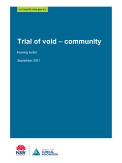
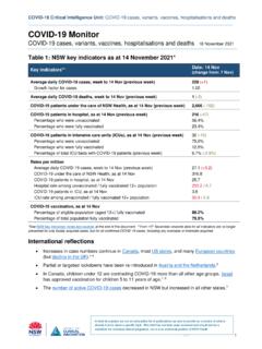
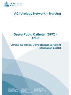
2 Figure 1: The Heart the heart is a cone-shaped, muscular organ located between the lungs behind the sternum. The heart muscle forms the myocardium the inner lining of the myocardium is called Endocardium and the outer layer cells is called the Epicardium. The pericardium (visceral) is the outer membranous sac with lubricating fluid. The heart has four chambers: two upper, thin-walled atria, and two lower, thick-walled ventricles. The ventricles are the chambers that eject blood in to arteries. The functions of the atrium are to receive the incoming blood from the vein LH_ICU2016_Learning_Package_Pacemaker_Le arning_Package 4|Page Liverpool Hospital Intensive Care: Learning packages Intensive Care Unit Pacemaker Learning Package The septum is a wall dividing the right and left sides of the heart Coronary arteries are the vessels that supply blood to the heart muscle. Cardiac valves : Cardiac valves permit blood to flow in only one direction through the heart Atrioventricular valves : 1] Tricuspid valve: separates the Right atrium from the Right ventricle 2] Bicuspid valve [Mitral valve]: lies between the Left atrium and Left ventricle.
3 Semi lunar valves : 1] pulmonic valve: is the valve between the Right ventricle and the pulmonary artery. 2] Aortic valve: is the valve between the Left ventricle and the aorta. The Heartbeat Each heartbeat is called a cardiac cycle. When the heart beats occur, the two atria contract together, then the two ventricles contract; then the whole heart relaxes. Systole is the contraction of heart chambers; diastole is their relaxation. The heart sounds, lub-dup, are due to the closing of the atrioventricular valves , followed by the closing of the semi lunar valves . Conduction System The hearts cardiac conduction functions by emitting electrical currents that cause the myocardium to contract. The atria are the first to contract then the ventricle. There are two specialized cells in the heart that keep the synchronization of the atria and the ventricle: 1. Nodal cells 2. Purkinje cells These cells have three characteristics that help to keep everything in time: 1.
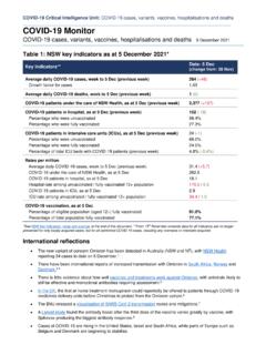
4 Automaticity: the ability to initiate an electrical impulse 2. Excitability: the ability to respond to an electrical impulse LH_ICU2016_Learning_Package_Pacemaker_Le arning_Package 5|Page Liverpool Hospital Intensive Care: Learning packages Intensive Care Unit Pacemaker Learning Package 3. Conductivity: the ability to transmit an electrical impulse from one cell to another. The electrical current is first initiated in the SA node, the hearts natural Pacemaker , located at the top of the right atrium. The SA node is composed of nodal cells. In a normal resting adult heart, the SA node initiates firing at 60 to100 impulses/minute, but the rate can change in response to the metabolic demands of the body. The impulses cause electrical stimulation and subsequent contraction of the atria. These signals then travel across the atrium to the atrioventricular node, located close to the septal leaflet of the tricuspid valve.
5 The AV node is also made up of nodal cells. The AV node coordinates these incoming electrical impulses. After a slight delay (giving the atria time to contract and complete ventricular filling), it relays the impulse to the ventricles. The Purkinje cells then take over in the ventricle. The impulse in the ventricles is initially conducted through a bundle of specialized conducting tissue called the bundle of His, which then divides into the right bundle branch (conducting impulses to the right ventricle). and the left bundle branch (conducting impulses to the left ventricle). To transmit impulses to the left ventricle, the heart's largest chamber, the left bundle branch divides into the left anterior and left posterior bundle branches. Impulses travel through the bundle branches to reach the Purkinje fibers (the terminal point in the conduction system). The Purkinje cells rapidly conduct the impulses through the thick walls of the ventricles.
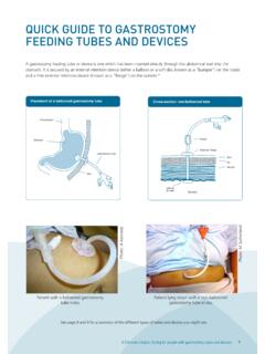
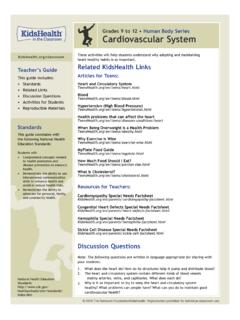
6 At this point, the myocardial cells are stimulated, causing ventricular contraction. (1). LH_ICU2016_Learning_Package_Pacemaker_Le arning_Package 6|Page Liverpool Hospital Intensive Care: Learning packages Intensive Care Unit Pacemaker Learning Package Figure 2: Cardiac conduction system. Marquette, Impulses can occur in various sites within the heart. Heart rate is determined by the Nodal and Purkinje cells with the fastest firing rate. Normally, the SA node has the highest inherent rate, 60 to100. impulses/minute. The AV node has an inherent rate of 40 to 60 impulses/minute Ventricular Pacemaker sites have the lowest inherent rate, of 30 to 40. impulses/minute. If the SA node malfunctions, the AV node generally takes over the heart's Pacemaker function at its inherently lower rate. Should both the SA and the AV nodes fail in their Pacemaker function, a Pacemaker site in the ventricle will fire at its inherent bradycardic rate of 30 to 40 impulses/minute.
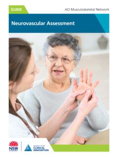
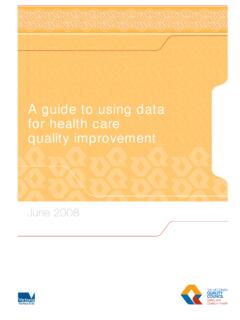
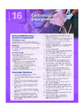
7 It is when one or both of these nodes fail that a temporary Pacemaker may be needed. Electrical activity of the heart is recorded on the ECG. LH_ICU2016_Learning_Package_Pacemaker_Le arning_Package 7|Page Liverpool Hospital Intensive Care: Learning packages Intensive Care Unit Pacemaker Learning Package Figure 3: Electrical representation and cardiac cycle. Marquette, LH_ICU2016_Learning_Package_Pacemaker_Le arning_Package 8|Page Liverpool Hospital Intensive Care: Learning packages Intensive Care Unit Pacemaker Learning Package DEFINITIONS. There are numerous terms used in pacing. Some of these are listed below: Action Potential: The action potential is brought on by a rapid change in membrane permeability to certain ions, with unique properties necessary for function of the electrical conduction system of the heart. Automaticity: Is the ability of the cardiac muscles to depolarize spontaneously, without external electrical stimulation from the nervous system.
8 Ampere (AMP, A): Is a measure of electrical current flowing past a point in a conductor when one volt of potential is applied across one ohm of resistance. In pacing, these currents are so small that they are expressed in thousandths of amperes (milliamperes, mA) or in millionths of amperes (microamperes, A). Amplitude: The maximum absolute value attained by an electrical waveform, voltage or current. The amplitudes of Pacemaker output pulses are expressed in volts (the difference in electrical potential), or in milliamperes (the measure of the electrical current flow). AV Interval / Atrioventricular Interval, AV Delay: In a dual chamber Pacemaker mode the AV Interval is the period of time between an atrial event (sensed or paced) and a scheduled paced ventricular event. It is typically measured in milliseconds. The AV Interval can also be thought of as the Pacemaker equivalent to the PR interval in normal conduction.
9 LH_ICU2016_Learning_Package_Pacemaker_Le arning_Package 9|Page Liverpool Hospital Intensive Care: Learning packages Intensive Care Unit Pacemaker Learning Package Asynchronous pacing: (a = not; syn = together; chrono = time) are those that are not together in time with the heart because they do not know what the heart is doing. Pacemakers such as AOO and VOO are asynchronous as they stimulate the heart at a fixed, preset rate independent of the electrical and/or mechanical activity of the heart. Burst Pacing: Is a technique of rapid pacing which may be used to terminate a tachycardia by delivering a series of pacing pulses in which the delivery rate remains constant within each pacing attempt. This technique is used in both the atria and the ventricles to attempt to terminate organized arrhythmias. Capture: Is initiation of depolarization of the atria and/or ventricles by an electrical stimulus delivered by an artificial Pacemaker .
10 Capture can be visualized on the monitor by a spike before every p wave (for atrial pacing) and a spike before every QRS (for ventricular pacing). Demand Pacing: A pacing stimulus is delivered to the myocardium if the intrinsic rate falls below the set rate on the Pacemaker . Dual-Chamber Pacemaker : A Pacemaker with two leads (one in the atrium and one in the ventricle) to allow pacing and/or sensing in both chambers of the heart to artificially restore the natural contraction sequence of the heart. Epicardial Pacing: Is a type of temporary pacing where pacing wires are fixed directly to the myocardium (ventricular and often atrial) and are exposed through the skin on the chest wall, usually following cardiac surgery. LH_ICU2016_Learning_Package_Pacemaker_Le arning_Package 10 | P a g e Liverpool Hospital Intensive Care: Learning packages Intensive Care Unit Pacemaker Learning Package Fixed Rate Pacing: A pacing stimulus is delivered to the myocardium at a programmed fixed rate regardless of the underlying rate and rhythm.