Transcription of Prenatal Detection of Congenital Heart Defects at the 11 ...
1 Prenatal Detection of Congenital HeartDefects at the 11- to 13-Week ScanUsing a Simple color doppler ProtocolIncluding the 4-Chamber and 3-Vesseland Trachea Viewsn the past years, plenty of reports about indirect screeningmethods for the diagnosis of Congenital Heart Defects werepublished. They were based on markers of aneuploidy,including nuchal translucency, triuspid regurgitation, and ductusvenosus velocimetric 4 However, the Detection ratesfor Congenital Heart Defects do not exceed 40% for nuchal translu-cency above the 95th percentile alone and 58% for nuchal translucencyabove the same limit combined with a reversed or absent a-wave inthe ductus venosus flow , indirect methods inMarcin Wiechec, MD, Anna Knafel, MD, PhD, Agnieszka Nocun, MDReceived January 31, 2014, from the Chair ofGynecology and Obstetrics, Jagiellonian Univer-sity, Krakow, Poland.
2 Revision requested March22, 2014. Revised manuscript accepted for publi-cation July 22, correspondence to Marcin Wiechec,MD, Chair of Gynecology and Obstetrics, Jagiellonian University, 23 Kopernik St, 31-501 Krakow, : 2015 by the American Institute of Ultrasound in Medicine |J Ultrasound Med 2015; 34:585 594|0278-4297 | RESEARCHO bjectives The first goal of this study was to analyze the diagnostic performance of the4-chamber view, 3-vessel and trachea view, and their combination in color mappingduring early cardiac evaluations for selecting cases suspicious of Congenital Heart second goal was to describe the most common abnormal flow patterns at thelevels of the 4-chamber and 3-vessel and trachea views in the late first We conducted a prospective observational study in which a simple cardiacsonographic protocol was applied in fetuses at gestational ages of 11 weeks to 13 weeks6 days.
3 Results A total of 1084 patients with known postnatal or autopsy findings wereincluded in the study. The median maternal age was years (range, 27 40 years).The median crown-rump length was mm (range, 45 84 mm). Overall, there were35 cases with a confirmed Congenital Heart defect ( ), including 16 accompaniedby aneuploidy. We found that our simple first-trimester cardiac protocol was an effec-tive screening method for Congenital Heart Defects . The most effective approach of the3 evaluated by us was the combined application of the 4-chamber and 3-vessel and tracheaviews in color mapping compared to the 4-chamber and 3-vessel and trachea views defined the most common ventricular inflow patterns and the V sign.
4 The techniquewe used was simple and easy to We confirmed that evaluation by two basic cardiac views allows for selec-tion of most cases with a univentricular Heart , atrioventricular septal Defects , coarctationof the aorta, pulmonary stenosis, pulmonary atresia, and conotruncal Defects . Key Words cardiac Defects ; fetal echocardiography; fetal Heart ; first trimester; first-trimester screening; obstetric ultrasound 1 3/4/15 3:13 PM Page 585the first trimester are at risk of false-positive findings due tothe functional immaturity of the cardiovascular system atthis stage of fetal development. It was discovered that tran-sient abnormal fetal cardiac flow patternscould be identifiedduring this the other hand, pulsed waveDoppler venous pulsatility index measurement protocolsare difficult to follow by nonexperienced examiners andare at risk of low reproducibility at primary fetal fetal echocardiography is generally definedas a fetal cardiac scan performed until 16 weeks ,8It is usually considered a highly specialized scanwhen the following specific indications are present.
5 Increased nuchal translucency measurement, congenitalheart defect risk factors, and early diagnosis of extracardiacfetal abnormalities. At our department, however, evalua-tion of the fetal Heart at the time of the nuchal translucencyscan has become a routine part of the late first-trimesterscan since 2007. According to the literature, this approachincreases the Detection rate of Congenital Heart Defects inthe first trimester to nearly 60% to 80%.8,9 Taking into account the performance of indirectscreening methods and the low availability of early fetalechocardiography, examiners with less experience expectsimple rules describing how to effectively select patientswith a high risk of Congenital Heart Defects based not onlyon the maternal history and secondary signs such as nuchaltranslucency and triuspid regurgitation but also on directbasic cardiac views.
6 The first goal of this study was to ana-lyze the diagnostic performance of the 4-chamber view,3-vessel and trachea view, their combination in color map-ping, and nuchal translucency during early cardiac evalua-tions for selecting cases of Congenital Heart Defects . Thesecond goal was to describe the most common abnormalpatterns of flow at the levels of the 4-chamber and 3-vesseland trachea views in the late first and MethodsThe study group included women who underwent sono-graphic examinations at gestational ages of 11 weeks and13 weeks 6 days at the ultrasound laboratory of the Chairof Gynecology and Obstetrics, Jagiellonian University,between January 2009 and June 2012.
7 The local EthicsCommittee approved the study protocol, and all partici-pating patients gave written consent. This group of whitepregnant women, who were prospectively examined, wentthrough detailed late first-trimester sonographic screeningfor aneuploidy, which was performed under condition thatthe fetal crown-rump length measured between 45 and 84mm and followed Fetal Medicine Foundation maternal body mass index was calculated in kilogramsper square meter on the day of the sonographic analysis was based on patients who were examinedwith Voluson E6 ultrasound scanner (GE Healthcare, Zipf,Austria) by experienced physicians ( , , and ).
8 The factory presets of the machine were modified to obtainimages of high diagnostic quality and to ensure adequatesafety limits for an early cardiac scan: the mechanical indexfor B-mode imaging and the soft tissue thermal index inthe color flow mode were set not to exceed a value of scans were performed with a transabdominal 4 8-MHzhybrid transducer. In difficult scanning conditions, atransvaginal 5 9-MHz hybrid transducer was above-mentioned standardized settings allowed foraccurate cardiac imaging and shortening of the scan dura-tion to fulfill the ALARA (as low as reasonably achievable) ,11 The fetal Heart examination followed a standardizedprotocol.
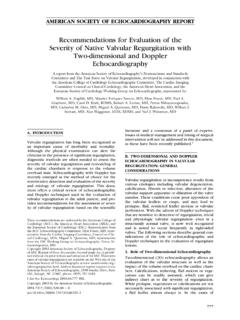
9 In all cases, an operator insonated the fetal chestso that the fetal spine was visualized at the 6- or 12-o clockposition to avoid oblique sections. Then color flow mappingwas activated for the shortest time possible. An idealinsonation angle to the interventricular septum of approxi-mately 45 guaranteed easy access to the levels of the 4-chamber and 3-vessel and trachea views, allowing for asmooth transition between these levels (Figure 1).At the level of the 4-chamber view in color mapping,the following criteria were assessed: number of stripes,subjective impression of their size ratio, and their separa-tion (interventricular septum).
10 At the level of the 3-ves-sel and trachea view in color mapping, the followingcriteria were evaluated: number of arterial arms, subjectiveWiechec et al color doppler Protocol for Prenatal Detection of Congenital Heart DefectsJ Ultrasound Med 2015; 34:585 594586 Figure 1. Ideal insonation of the beam (45 to the interventricular septumwith the fetal spine at the 6-o clock position) for an early cardiac evalua-tion starting from the ventricular inflows at the level of 4-chamber viewthrough the 3-vessel view and finishing with depiction of the V sign atthe level of the 3-vessel and trachea 1 3/4/15 3:13 PM Page 586impression of their size ratio, and flow direction.