Search results with tag "Color doppler imaging"
Efficient Implementation of Ultrasound Color Doppler ...
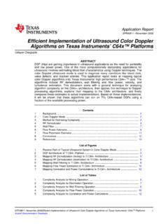
www.ti.com1 Background 2 Color Doppler Mode ransducer Frontend + RX Beamformer B-Mode Imaging Color Scan Converter Color Doppler Imaging Tissue/ Flow Decision Color
Recommendations for Quantification of Doppler ...
asecho.orgwave (CW) Doppler,and color Doppler imaging.PW Doppler measures flow velocity within a specific site (or sample volume) but is limited by the aliasing ... Recommendations for Quantification of Doppler Echocardiography: ... color flow Doppler can help determine the direction

Peripheral Arterial Ultrasound Examinations Using Color ...
www.aium.orgThe sonographic examination consists of grayscale/color Doppler imaging and spectral Doppler waveforms in the appropriate arterial segments. Color Doppler imaging should be ... tion of blood flow and the direction of the ultrasound beam is kept at 60° or less. Velocity esti-

Role of Color Flow Ultrasound in Detection of Deep Venous ...
files.eric.ed.govB-mode and color Doppler imaging is needed for early diagnosis of DVT to prevent complications and squeal of DVT.Aim and objectives: the objectives of our study were to evaluate the role of color flow Doppler in the diagnosis of deep vein thrombosis (DVT), and subsequently to …

Benign Mimics of Malignancy on Breast Imaging
www.umassmed.eduUltrasound: Echogenic solid mass with marked vascular flow on color Doppler imaging at 8 o'clock 10 cm from the nipple measuring 6 x 4 x 6 mm, corresponding to the site of the mammographic mass. Pathology: This benign vascular lesion consists of a proliferation of