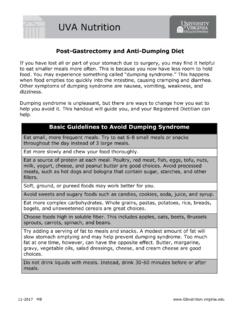
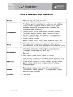
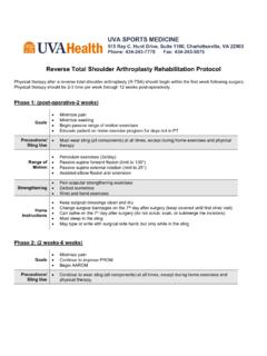
Transcription of Prevention and Management of Complications of …
1 PRACTICAL GASTROENTEROLOGY NOVEMBER 200466 INTRODUCTIONS ince its introduction in 1980 (1), the use of per-cutaneous endoscopic gastrostomy (PEG) tubeshas increased exponentially. While an estimated61,000 PEG tubes were placed in 1988, an estimated216,000 are performed annually today, making PEGplacement the second most common indication forendoscopy of the upper gastrointestinal tract (2). Up to10% of nursing home residents and as many as Medicare patients over the age of 85 years undergogastrostomy (3). As data demonstrating the benefits ofenteral over parenteral nutrition mounts, and ourelderly population grows, we can expect the use ofPEG tubes to continue to rise. However, the placementof a PEG tube is not without its risks. The overall com-plication rate has remained stable over the last 15-20years, ranging from 4% to of cases (4 7). Threeto 4% of all cases are affected by major Complications , those that are life threatening and/or require surgi-cal intervention or hospitalization (Table 1) (4,6,8).
2 The more common minor Complications occur inbetween and of cases (Table 2) (4,6,9). Prevention and Management of Complications of PercutaneousEndoscopic Gastrostomy (PEG) TubesNUTRITION ISSUES IN GASTROENTEROLOGY, SERIES #22 Carol Rees Parrish, , MS,Series EditorChristopher R. Lynch, , Department of InternalMedicine, University of Utah School of Medicine, SaltLake City, Utah. John C. Fang, , Associate Pro-fessor of Medicine, Division of Gastroenterology,Department of Internal Medicine, University of UtahSchool of Medicine, Salt Lake City, number of patients with PEG tubes continues to rise, and coincident with that rise,more gastroenterologists are being consulted with Complications of PEG of PEG tube Complications are minor, but several have the potential to causesignificant morbidity and even mortality if not recognized and managed and early identification of PEG Complications will reduce morbidity and costsubstantially.
3 Expertise in the Management of these Complications is critical to the prac-ticing R. Lynch(continued on page 68)John C. FangPRACTICAL GASTROENTEROLOGY NOVEMBER 200468 NUTRITION ISSUES IN GASTROENTEROLOGY, SERIES #22 Prevention and Management of Complications of PEG TubesWhile the overall mortality post-PEG placement ishigh due to underlying co-morbidity, the rate of proce-dure-related mortality and 30-day mortality attribut-able to PEG placement itself are extremely low (0% to2% and to respectively) (6,10 12). Itshould be noted that mortality associated with PEGplacement is significantly higher in hospitalizedpatients (13), patients with diabetes, poor nutritionalstatus, and long-term corticosteroid administration (8).Complication rates of percutaneous gastrostomy tubesplaced endoscopically versus radiologically using flu-oroscopy are similar (14,15). Enteral nutritional support is indicated for patientswith poor volitional intake, permanent neurologicalimpairment, oropharyngeal dysfunction, short gut syn-drome, and major trauma and burns (16).
4 Generallypatients who meet one or more of these criteria formore than 30 days are candidates for PEG placement. Absolute contraindications to PEG placement arethe same as those of upper gastrointestinal endoscopyas well as an inability to transilluminate the abdominalwall and appose the anterior gastric wall. Relative con-traindications include coagulopathy, gastric varices,morbid obesity, prior gastric surgery, ascites, chronicambulatory peritoneal dialysis (CAPD), and neoplas-tic, infiltrative, or inflammatory disease of the gastricor abdominal wall (17).PROCEDURE-RELATED COMPLICATIONSThe overall success rates for PEG placement are con-sistently reported at 94% to 98% (4,18,19) and com-pare favorably with fluoroscopic placement by a radi-ologist (18,20). The pull and push techniques result insimilar success rates (21). Factors that can lead tounsuccessful PEG placement can include obstructionof pharynx or esophagus, deterioration of the clinicalstatus of the patient intraprocedurally, poor transillu-mination of the abdominal wall, incidental finding ofgastric cancer, and development of hematoma at thegastrostomy site (4).
5 Prior surgery that results in alter-ation of esophageal or gastric anatomy can also lead toa difficult PEG placement (22).Patients undergoing PEG tube placement are sub-ject to the Complications associated with upperendoscopy and sedation. While the rate is low ( ),significant morbidity can result from these complica-tions; the most common Complications of endoscopyinclude perforation, hemorrhage, and aspiration (23),while sedation carries the risks of hypoxia, aspiration,and hypotension (24,25). It is not documented, but therisks of sedation are likely higher in the more severelydebilitated PEG population. AspirationUpper gastrointestinal endoscopy is associated with asignificant risk of aspiration. In a report in which 15%of 64 patients had aspiration related to PEG placement,only 2 of the patients had aspiration during the proce-dure while the other 11 did so over the next severalweeks for reasons unrelated to PEG placement (26).
6 Inother reports, aspiration related to the procedure itselfoccurred in to of cases (4,27). Risk factorsfor intra-procedural aspiration include supine position,sedation, neurological impairment, and advanced age(17). The endoscopist can minimize the risk of thiscomplication by avoiding over-sedation, minimizingair insufflation of the stomach, thoroughly aspirating(continued from page 66)Table 1 Major , 27 Hemorrhage0% , 29, , 5 Necrotizing fasciitis rare50 53 Death0% , 10 12 Tumor implantationrare67 70 Table 2 Minor ComplicationsComplicationFrequencyRefere ncesIleus1% 2%4, 27 Peristomal 30%39 41 Stomal leakage1% 2%54 Buried , 56, 57 Gastric , 29, 31, 60 Fistulous , 60, 61the gastric contents before the procedure, and perform-ing the procedure efficiently (17). Demortier, et al (28)have reported promising results using an unsedatedtransnasal approach to PEG placement, using a small-diameter endoscope, to lower the risks of bleeding during PEG placement is an uncommoncomplication, occurring in approximately 1% of cases(5,29,30).
7 A review of 1338 patients reported that lessthan of cases are complicated by hemorrhagerequiring transfusion and/or laparotomy (31). Risk fac-tors include anticoagulation and previous anatomic alter-ation (32). A case of fatal retroperitoneal hemorrhagebelieved to be associated with surgically altered anatomyhas been reported (33). The development of a hematomaat the PEG site complicates roughly 1% of cases (5).Perforation of Viscera/PeritonitisComplete laceration of the stomach, small bowel, orcolon is a potentially catastrophic complication occur-ring in to of cases (4,5). It is recognized,however, that transient subclinical pneumoperitoneumoccurs during PEG placement in as many as 56% ofprocedures and is generally not of any clinical signifi-cance (34). Peritonitis, manifested in the post-PEGpatient as abdominal pain, leukocytosis, ileus, andfever, can result in significant morbidity if not identi-fied and treated early (35).
8 The prevalence of persis-tent subclinical pneumoperitoneum limits the utility ofplain films for evaluation of suspected fluoroscopic imaging of the PEG tube withinfusion of water-soluble contrast is most useful toevaluate visceral integrity in patients in whom peri-tonitis is a consideration (36). If active leakage of con-trast is identified in a patient with clinical signs of peri-tonitis, broad-spectrum antibiotics and surgical explo-ration are usually IleusIt has been established that tube feedings may begin assoon as 3 hours after PEG placement (37). However, in1% 2% of cases prolonged ileus may follow PEGplacement, and should be managed conservatively (4).Acute gastric distension post-PEG placement can bedecompressed by simply uncapping the PEG tube (38).POST-PROCEDURE COMPLICATIONSThe PEG site should be cleaned with mild soap andwater hydrogen peroxide should not be used as it canirritate the skin and contribute to stomal leaks.
9 Cut drainsponges should be placed over, rather than under, theexternal bumper so as not to apply excessive tension tothe PEG site. Occlusive dressings should not be used asthey can lead to peristomal skin maceration and break-down. Should excessive granulation tissue develop atthe PEG site, topical silver nitrate can be applied toreduce irritation and decrease drainage (Figure 1). PEG Site InfectionThe most common complication of PEG placement isinfection at the PEG site. As many as 30% of cases arecomplicated by peristomal wound infection (39 41),however more than 70% of these are minor with lessthan of stomal infections requiring aggressivemedical and/or surgical treatment (42). Patients withdiabetes, obesity, poor nutritional status, and those onchronic corticosteroid therapy are at increased risk forinfection (43). Excessive pressure between the PEG sexternal and internal bolsters is associated with ahigher infection rate thus setting and maintaining theproper tension can decrease the likelihood of contact of the outer bolster with the skin is allthat is required to appose the gastric and abdominalwall.
10 The introducer technique that does not pull thePEG tube through the oropharynx has been shown toresult in fewer infections compared to the pull or pushtechniques (44,45). The administration of prophylactic antibioticsprior to PEG placement reduces the risk of trials have demonstrated the benefit of a sin-gle, broad-spectrum antibiotic immediately prior toPEG placement (42,46 48). The use of prophylacticantibiotics is cost-effective as well (49). It is generalpractice to administer a single dose of a first or thirdgeneration cephalosporin 30 minutes prior to the pro-cedure. Prophylaxis is not necessary in those patientsalready receiving comparable antibiotics for other rea-PRACTICAL GASTROENTEROLOGY NOVEMBER 200469 NUTRITION ISSUES IN GASTROENTEROLOGY, SERIES #22 Prevention and Management of Complications of PEG Tubessons at the time of PEG placement. An adequate skinincision, 1 2 mm larger than the feeding tube, whichcan allow egress of bacteria and gastric secretions,may also reduce infection risk.