Transcription of Reticulocyte Count - Pearson Education
1 Reticulocyte CountPrincipleUsing a supravital stain ( , new methylene blue), residual ribosomal RNA within the reticulocytes is precipitated. An equal volume of stain is added to EDTA-anticoagulated blood, the dilution mixture incubated, and a smear is prepared. The smear is examined to determine the number of reticulocytes present. An erythrocyte containing two or more particles of blue-stained material is a Reticulocyte . The number of reticulocytes is expressed as a percentage of the total number of erythrocytes and Equipment1. New methylene blue N New methylene blue N g(certified by Biological Stain Commission)NaCl gQS with distilled water to 100 mLFilter staining solution through Whatman No. 1 filter paper prior to use2. Test tubes, 12 x 75 mm3. Pasteur pipets4. Glass slides5. Spreader slide6. Microscope7. Miller ocular disc8. Hand counterQuality ControlCommercial quality control materials with established control limits should be run periodically.
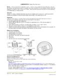
2 The frequency is determined by each laboratory's workload. For instance, quality control material may be run at the beginning of each eight-hour Whole blood anticoagulated with EDTA is recommended; however, any anticoagulant is acceptable. Free-flowing capillary blood may be Reticulocyte Staining:a. Pipet five drops of new methylene blue N solution into a labeled 12 x 75 mm test Add an equal volume (five drops) of well-mixed blood (control or patient).c. Mix gently using a pasteur Incubate at room temperature for 10 minutes. 2. Resuspend mixture thoroughly, and prepare 2-3 wedge smears. Air-dry the smears immediately by gently waving the Label the Reticulocyte smears by writing appropriate information directly into the thick end of the Reticulocyte Counting:a. Using the oil immersion lens and an ocular fitted with a Miller disc (Figure 7-11), select an area of the smear where the erythrocytes are evenly distributed, without overlapping.
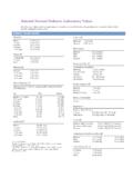
3 There should be 80-100 erythrocytes per oil immersion Count a total of 20 fields. In each field, Count erythrocytes (include reticulocytes) in the smaller square (B) and reticulocytes in the larger square (A). 5. A second clinical laboratory professional should perform a Reticulocyte Count on a second smear in the same The calculation formula for determining the percentage of reticulocytes present is given in Web Figure 7-6. Reticulocyte Count (%) = Total # erythrocytes counted (Square B) X 9 X 100 Total # reticulocytes counted (Square A) 2. The absolute Reticulocyte Count is determined by the following calculation:Absolute Reticulocyte Count = Total erythrocyte Count (x10 /L) x Reticulocyte %12 For example: If the Reticulocyte Count is 1% and the erythrocyte Count is 5 x 10 /L:121% x (5 x 10 /L) = .05 x 10 /L or 50 x 10 /L12129 Reference Interval RelativeAbsoluteAdult - x 10 /L9 Adult - %18-158 x 10 /L9 - x 10 /L9 Comments1.
4 Allowing the incubation time to exceed 15 minutes increases the possibility of erroneous results due to the dye adhering to mature The degree of imprecision between Reticulocyte counts performed on the same blood sample is dependent on the total number of reticulocytes and the individual or individuals who perform the counts. If the Reticulocyte Count is within the reference interval, the error may be as great as 25%, whereas the error associated with a Reticulocyte Count of 5% or greater may be 10%. In general, the error will decrease as the uncorrected Reticulocyte Count Technical sources of error:a. Other RBC inclusions (Pappenheimer bodies, Howell-Jolly bodies, and Heinz bodies) will be stained with new methylene blue. Howell-Jolly bodies and Heinz bodies may be distinguished from precipitated reticulum by their shape and staining characteristics. Heinz bodies appear as light blue-green inclusion located at the periphery of the erythrocyte.
5 Howell-Jolly bodies are usually one or two round, deep purple staining inclusions and are also visible on Romanosky stains. Pappenheimer bodies are indistinguishable from reticulum of reticulocytes. If Pappenheimer bodies are suspected, a Prussian blue iron stain should be performed to verify their The whole blood-stain mixture should be resuspended prior to making the smears. Reticulocytes have a lower density than mature erythrocytes, and therefore will be located near the top during Poor drying or moisture may result in the presence of refractive artifact on the smears. This refractive artifact may be confused with precipitated reticulum. However, precipitated reticulum is not refractive, and fine focus adjustment will reveal the difference. d. Increased glucose levels may cause the reticulocytes to have a pale Counterstaining with a Romanosky-type stain is no longer recommended, as it may obscure the precipitated Misinterpretations may result when reporting only the percentage of reticulocytes present in the peripheral blood, because the Reticulocyte result is dependent on the total number of erythrocytes present in the peripheral blood.
6 If the total erythrocyte Count is decreased, the Reticulocyte percentage does not accurately reflect the bone marrow's production of new erythrocytes. The correction formulas ( , corrected Reticulocyte Count and Reticulocyte production index) used to avoid interpretation errors due to the total erythrocyte Count and increased bone marrow stimulation are discussed in detail in chapter ,4,55. A Reticulocyte production index greater than 3 is associated with hemolytic anemias ( , hereditary spherocytosis), recent hemorrhage and response to therapy. A Reticulocyte production index less than 2 is associated with hypoproliferative disorders ( , aplastic anemia) and ineffective erythropoiesis seen in megaloblastic National Committee for Clinical Laboratory Standards. Methods for Reticulocyte counting (flow cytometry and supravital dyes). H44-A. Villanova, Pa.: NCCLS; Gilmer PR, Koepke JA. The Reticulocyte : An Approach to Definition.
7 Am J Clin Pathol. 1976; 66 Peebles DA, Hochberg A, Clark TD. Analysis of manual Reticulocyte counting. Am J Clin Pathol. 1981; 76 Savage RA, Skoog DP, Rabinovitch A. Analytical Inaccuracy and Imprecision of Reticulocyte Counting: A Preliminary Report from the College of American Pathologists Reticulocyte Project. Blood Cells. 1985; 11 Hillman RS, Finch CA. Red Cell Manual. 5th ed. Philadelphia: Davis Co.; 1995-2003 by A Pearson Company Prentice-Hall, Notic