Transcription of Retrograde Intrarenal Surgery (RIRS) - …
1 OCTOBER 2009 IntroductionEver since the first ureteroscopy performed by HughHampton Young at the beginning of last century,1ureterorenoscopy has evolved over nearly a centuryfrom the era of happenstance to the Digital & Roboticera. This evolution culminates in the technique ofretrograde Intrarenal Surgery ( rirs ) utilising flexibleureterorenoscope and Holmium laser, which is a veryeffective treatment option in the Urologist'sarmamentarium when first-line treatment options, viz, extracorporeal shockwave lithtropsy (ESWL) andpercutaneous nephrolithotomy (PCNL) failed; it wouldalso be used as first-line treatment in selected first ureteroscopy began by Young, the "Father of Modern Urology",introduced a Fr 12 Paediatric cystoscope into themassively dilated ureter of a child with posteriorurethral valve in 1912.
2 He was able to advance up torenal pelvis and became the first Urologist to view theintrarenal collecting system of a patient enthusiasm for ureteroscopy cooled down until thefirst breakthrough in the late 1950's with thedevelopment of the first fibreoptic endoscope. The firstflexible ureterorenoscopy was performed by Marshallvia an ureterotomy using a Fr 9 flexible endoscope fordiagnostic purposes in then it was not until1977 that Goodman reported the first rigid ureteroscopyfor therapeutic purposes1; whereas Fuchs & Fuchs(1990) reported the first large series (208 patients) ofrenal calculi treated by flexible for contemporary rirs using flexibleureterorenoscope and Holmium laser for treatment ofrenal stones was set by Grasso & Chalik in rirs for Renal StonesInstrumentsStandard instruments for rirs include:Flexible ureterorenoscopeHolmium: yttrium-aluminium-garnet (YAG) laserVideo cameraFluoroscopic supportAccessory instrumentsGuide-wiresDilatorsAccess sheathBasketUreteric cathetersFlexible UreterorenoscopeStandard fibreoptic flexible ureterorenoscopes have atip size in the range of - 9Fr.
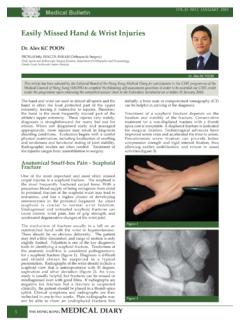
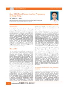
3 They are activelydeflectable (primary deflection) with 120 to 170 degreesof deflection in one direction and 170 to 270 degrees inthe other. Secondary deflection will be passive or active(Fig. 1); active secondary deflection allows bettermanoeuvrability especially in the lower pole have working channels of Fr - 4 and standardinstruments ( baskets) are Fr : YAG LaserThe Holmium:YAG laser is the lithotripter of choice forRIRS nowadays. It has a wavelength of 2100nm andtissue penetration of The laser energy isdelivered via quartz fibres to the stone surface, where itis absorbed and turned into heat energy that pulverisesthe stone into dust by a "photothermal" effect. Thus,stone fragment retrieval with basket / grasping forcepswould not be necessary. Laser lithotripsy can be carriedout safely in patients on sizeRetrograde Intrarenal Surgery ( rirs ) -Ureterorenoscopic lithotripsy for Renal StonesDr.
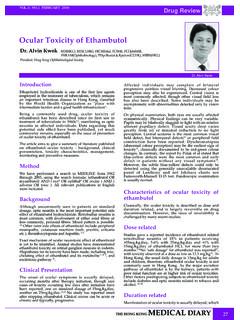
4 Wing-hang AUDr. Wing-hang AUAssociate Consultant UrologistDivision of Urology, Department of Surgery , Queen Mary & Tung Wah Hospitals,Li Ka Shing Faculty of Medicine, The University of Hong KongMBBS(HK), FRCS(Edin), FCSHK, FRCSEd(Urol), FHKAM( Surgery )Fig. 1a Flexible ureterorenoscope with active primary &passive secondary 1b Flexible ureterorenoscope with active primary &secondary MAY OCTOBER 2009200micron laser fibre used in flexible ureterorenoscopywill minimise the hindrance to scope deflection (Fig 2).The usual laser setting on commencing the procedurewould be Camera and FluoroscopyAccurate and clear visualisation of the ureter and pelvi-calyceal system using video camera is essential to thesuccess of the procedure. Delineation of the pelvi-calyceal anatomy using fluoroscopy and retrogradepyelogram helps the surgeon to orientate him/herselfthroughout the double flexible-tips guide-wire is essential forscope introduction in order to avoid damage to theflexible ureterorenoscope.
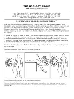
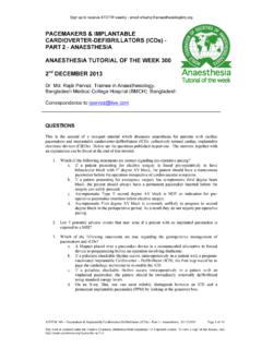
5 Standard PTFE-coated guide-wires would be used for the placementof ureteric catheters, introducing dilators / Accesssheath and serve as safety guide-wire (Fig. 3a).Dilators and Access SheathThe use of Access sheath (Fig. 3b) is optional duringRIRS, which would depend on the personalpreference of the surgeon, stone load and pelvi-calyceal anatomy. The size of commonly usedAccess sheathes include Fr 9/11 and Fr 12/14. SerialTeflon dilators up to size 16 would be used forureteral calibration and dilatation beforeintroducing the Access sheath over guide-wireunder fluoroscopic using an Access sheath include:Facilitate repeated introduction and withdrawalof the endoscope which would be required forpatients with large stone burden;Avoid build-up of pressure within the pelvi-calyceal system especially when pressurisedirrigating fluid is used to improve 2 Holmium: YAG laser machine and laser : 200micron laser fibre is used in rirs to minimise hindrance to 3a Guide-wires(Left: double flexible-tips; right: PTFE-coated)Baskets / ExtractorsDevelopment of the tipless Nitinol basket is vital forthe success of rirs of renal stones (Fig.)
6 3c). Tiplessdesign avoids traumatisation of the mucosa duringintrarenal manipulations. Nitinol baskets alsopreserve tip deflection of the flexibleureterorenoscope. Relocation of lower pole stonesinto renal pelvis or upper pole calyx with basketwill greatly enhance the efficiency of stonefragmentation. Extraction of stone fragments via theAccess sheath would be considered in patients withlarge stone burden. Latest designs ( NGate fromCooks Medical) allow a "frontal attack" to the stonesinstead of the usual sideway stone CathetersStandard Fr 6 / Fr 7 open-end ureteric catheters willbe used for Retrograde pyelogram at the beginningof the procedure and temporary drainage post-operatively. Fr 6 / Fr 7 double-J ureteric catheterswill be used for pre-stenting or temporary with double-J ureteric catheter forabout 2 to 4 weeks for ureteric dilatation wouldbe required to facilitate insertion of Accesssheath especially for oriental patients with a lessspacious ureter;Traumatisation of the ureter may occur duringureteral dilatation and introduction of the using an Access sheath include:Fig.
7 3b Ureteric access sheathFig. 3c Tipless nitinol OCTOBER 2009 IndicationsIndications of rirs for renal stones are listed as follows:1. Failed extracorporeal shockwave lithotripsy2. Radiolucent stones3. Concomitant ureteric and renal stones4. Anatomical problems infundibular stenosis5. Nephrocalcinosis6. Bleeding disorders57. Need for complete stone removal pilotPre-operative Assessment and ProcedureImaging assessments on stone load, stone location andpelvi-calyceal anatomy are essential before urogram is the mostcommonly used imaging modality. Pre-operativeretrograde pyelogram would be required for patientswith impaired renal function. CT urogram is becomingmore and more popular nowadays. An informedconsent should be obtained including counselling ontreatment options, procedure and potentialcomplications, whereby possibilities of requiring post-op stenting, second-look procedure, auxiliaryprocedure and failed procedure are all thoroughlyexplained.
8 Urine cultures are performed to ensure thatpatients have sterile urine before the with asymptomatic persistent bacteriuriashould be given an appropriate antibiotic forprophylaxis. Patients are put under generalanaesthesia with prophylactic antibiotics administeredon-induction. With the patient in Lloyd-Davisposition, cystoscopy and Retrograde pyelogram (RP)are performed to delineate upper tract anatomy andany stone migration before instrumentation is points of rirs :Safety guide-wire inserted up to renal pelvis;Ureteric dilatation and use of Access sheath aspreferred / indicated;Flexible ureterorenoscope "rail-roaded" up to renalpelvis over double-flexible tips guide-wire underfluoroscopic and endoscopic guidance;Systematic inspection of the pelvi-calyceal system toidentify pathology endoscopically under salineirrigation (pressurised irrigant as required,preferably with Access sheath) and aided withfluoroscopy / RP as required;Commence lithotripsy with Holmium laser;Stone relocation / retrieval with basket as indicated.
9 Assess stone clearance with endoscopy / fluoroscopy/ RP;Placement of double-J ureteric catheter as will usually have their first follow-up visitscheduled about 2 weeks post-op. Treatment outcomewill be assessed with a KUB radio-graph or additionalimaging as of rirs for Renal StonesMichael Grasso was credited for setting the benchmarkof contemporary rirs for renal stones. 228 patients weretreated with rirs in his series reported in rate (stone fragment <2mm in size) was 81% afterprimary rirs and improved to 90% after secondaryRIRS. Best results were achieved for upper & mid-polestones with 90% success rate after primary treatment andup to 97% success after secondary rirs . Grasso &Ficazzola have reported their results of rirs in treatinglower pole renal rirs were performed in 79patients.
10 Patients were analysed in 3 groups with stonesizes of 10mm or less (group 1), 20mm or less (group 2)and more than 20mm (group 3). The overall completefragmentation rate (stone fragment <2mm in size) was91%. Complete fragmentation rate was 94% and 95% forgroup 1 and 2 respectively after one session; it was 45%after one session and 82% after two sessions for group excellent results of Grasso inspired the Lower Pole IIstudy, which was a prospective randomised studyinvolving 19 with lower pole renalstones of 1cm or less were randomised to receive ESWLor flexible ureterorenoscopy (URS). 78 patients wererandomised in total and 67 patients remained onprotocol. Treatment outcome was assessed by non-contrast CT. Stone-free rates at 4 months follow-up were35% and 50% for ESWL and flexible URS respectively,but the difference was not statistically significant.