Transcription of Samples: Chapter 05
1 Overview of the RegionBONES AND BONE MARKINGSOF THE REGION Pelvic GirdleFemur PatellaTibia and FibulaBones of the FootJOINTS, LIGAMENTS, ANDBURSAE OF THE REGIONS acroiliac JointHip JointKnee JointTibiofibular JointsAnkle JointsJoints That Permit Inversion and EversionRemaining Joints Within the FootCONNECTIVE TISSUESTRUCTURES OF THE REGIONDeep Investing Fascia and Iliotibial BandFascial Compartment Divisions in the LegPlantar Fascia or AponeurosisIndividual MusclesHip Joint MusclesPIRIFORMIS AND THE OTHERDEEP LATERAL ROTATORS OFTHE HIPADDUCTOR MAGNUSADDUCTOR LONGUS AND BREVISPECTINEUSGRACILISGLUTEUS MINIMUS GLUTEUS MEDIUSTENSOR FASCIA LATAEGLUTEUS MAXIMUSHAMSTRINGS:SEMIMEMBRANOSUSHAMSTRI NGS:SEMITENDINOSUSHAMSTRINGS: BICEPSFEMORISILIOPSOAS (PSOAS MAJOR ANDILIACUS)QUADRICEPS GROUP: VASTUSINTERMEDIUSQUADRICEPS GROUP: VASTUSMEDIALISQUADRICEPS GROUP: VASTUSLATERALISQUADRICEPS GROUP: RECTUSFEMORISSARTORIUSR egional Illustrations of MusclesPosterior Knee and Superficial Posterior Leg MusclesPOPLITEUSPLANTARISGASTROCNEMIUSSO LEUSM uscles of the Leg That Movethe Foot and ToesTIBIALIS POSTERIORFLEXOR DIGITORUM LONGUSFLEXOR HALLUCIS LONGUSPERONEUS LONGUSPERONEUS BREVISPERONEUS TERTIUSEXTENSOR DIGITORUM LONGUSEXTENSOR HALLUCIS LONGUS Chapter OUTLINEL ower Limb5(Continued)297 LWBK788-Ch5_297-437_Moorecraft Edition 1 24/01/11 9:24 PM Page 297298 PART 2 INDIVIDUAL MUSCLES BY BODY REGIONOVERVIEW OF THE REGIONThe lower limb is designed for weight-bearing, balance, andmobility.
2 The bones and muscles of the lower limb are largerand stronger than those of the upper limb, which is necessaryfor the functions of weight-bearing and balance. Our lowerlimbs carry us, allow us to push forward, and also keep usstanding still. Our sense of steadiness and strength oftencomes from our lower limbs. The muscles of the thigh are thick and strong and can tol-erate greater pressure during massage than the smaller mus-cles of the arm. P trissage is generally welcome and easy toperform in the thigh. The muscles of the posterior leg arealso thick and strong, as they propel us forward. The ante-rior leg is less muscular and more suited to friction or deepeffleurage. The foot is our anchor, grounding us to the earth. Althoughcomposed of a complex structure of bones, joints, and mus-cles, the foot is also our steady connection to the AND BONE MARKINGS OF THE REGIONThe bones of the lower limb include the pelvic girdle, femur,patella, tibia, fibula, and bones of the foot.
3 These are dis-cussed GirdleThe pelvic girdle contains the hip bone and the sacrum. As al-ready noted, the hip bone contains the ilium, ischium, andpubis (see Chapter 4). Recall that the iliac crest contains theanterior superior iliac spine (ASIS) and the posterior supe-rior iliac spine (PSIS). The iliac spine contains the entire iliaccrest and extends inferiorly in the front and back to includethe anterior inferior iliac spine (AIIS) and posterior inferioriliac spine (PIIS), as well. The anterior aspect of the ilium isbroad and curved, like a fossa. It is called the iliac fossa. Recall that the ischium has a significant bone marking inthe ischial tuberosity. The hamstring muscles connect to the -ischial tuberosity. In addition, the ischium contains a spine,which separates the greater sciatic notch from the lesser that the pubis contains two rami, the superiorpubic ramus and the inferior pubic ramus.
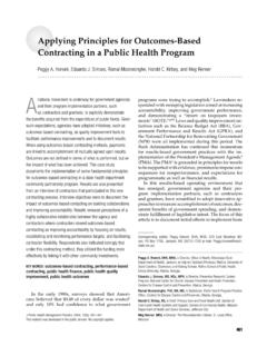
4 The thigh adduc-tors originate on the that the acetabulum is the name of the socket thatarticulates with the head of the femur to form the hip acetabulum is where the ilium, ischium, and pubis jointogether. Figure 5-1 shows bones and bone markings of femur, or thigh bone, is the longest and strongest bonein the body. Its rounded head, located on the proximal, me-dial aspect of the femur, fits beautifully in the acetabulum toform the hip joint. The greater trochanter is a sizable bonemarking on the lateral aspect of the proximal femur. Thelesser trochanter is smaller and is located distal and slightlyposterior to the head of the femur on the medial aspect of thebone. Rounded medial and lateral condyles are located onthe distal end of the femur and articulate with the tibia. Arough line called the linea asperaruns almost the full lengthof the posterior femur.
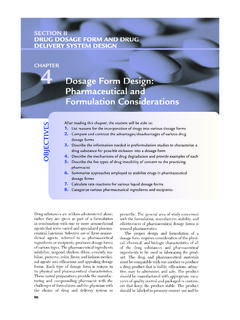
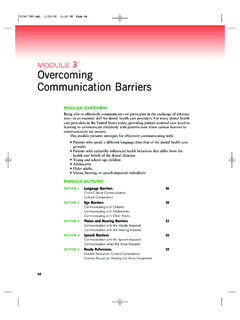
5 The gluteal tuberosity is located onthe proximal, posterior femur, very close to the proximal lineaaspera. The pectineal line is located proximal and medial onthe posterior femur, just inferior to the lesser 5-2 illustrates the femur and its bone markings, as wellas the patella. PatellaThe patella or knee cap is a sesamoid bone that lies anteriorto the junction of the femur and tibia. The patella is cartilagi-nous at birth and ossifies between 3 and 6 years of age. Thepatella is embedded in the quadriceps tendon and causesthe tendon to be positioned more anteriorly, thus enhanc-ing the leverage of the quadriceps tendon as it pulls on thetibial tuberosity to extend the knee. The patella slides up andTIBIALIS ANTERIORR egional Illustrations of MusclesIntrinsic Foot MusclesDORSAL INTEROSSEIPLANTAR INTEROSSEILAYER 3 INTRINSIC FOOTMUSCLESFLEXOR HALLUCIS BREVISADDUCTOR HALLUCISFLEXOR DIGITI MINIMI BREVISLUMBRICALSQUADRATUS PLANTAEABDUCTOR HALLUCISFLEXOR DIGITORUM BREVISABDUCTOR DIGITI MINIMIR egional Illustrations of MusclesILLUSTRATIONS OF NERVESUPPLY AND ARTERIAL SUPPLYTO LOWER LIMBC hapter SummaryWorkbook Muscle Drawing ExercisesPalpation ExercisesClay Work ExercisesCase Study ExercisesReview ExercisesLWBK788-Ch5_297-438:Moorecraft Edition 1 1/29/11 4:27 PM Page 298down as we flex and extend the leg.
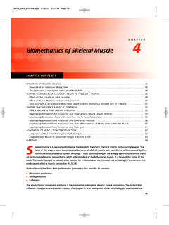
6 Cartilage on the poste-rior aspect of the patella provides cushioning between thepatella and the femur (see Fig. 5-2).Tibia and FibulaThe tibia and fibula are the bones of the leg. The tibia ismuch the larger and is located medial to the fibula. The tibiais the weight-bearing bone and is part of the knee joint. Sev-eral important bone markings exist on the tibia and proximal end of the tibia contains two condyles, a me-dial condyle and a lateral condyle. The tibial tuberosity is lo-cated on the proximal anterior aspect of the tibia, justinferior to the patella. As already noted, it serves as the in-sertion site for the quadriceps tendon. Pes anserinus, whichCHAPTER 5 Lower Limb299 AIliac crestIliac fossaAnterior superioriliac spineAnterior inferioriliac spineIschiumPubisIliumThe pubic symphysis is themedial junction between theright and left sides of thepelvic girdle.
7 It is a slightlymoveable cartilaginous joint. L5L5 Posterior superioriliac spinePosterior inferioriliac spineSacrumIschial tuberosityCoccyxIliumThe sacroiliac joint is a large,stable union between the lateralsacrum and medial ilium. BFIGURE 5-1 Bones and bonemarkings of the pelvis A: Anteriorview; B: Posterior view; C: Lateralviewmeans goose foot, is the name given to a flat area on theproximal, anterior, medial tibia, just medial to the tibialtuberosity. Three muscles insert at pes anserinus, and thetriplet of tendons looks somewhat like the three toes of agoose s foot. On the distal medial side of the tibia is the me-dial malleolus, which is commonly referred to the innerankle bone in lay terms. The fibula contains some important bone markings, aswell. The head of the fibula is the bone s most proximal as-pect. Two important muscles connect to this bone , the fibula has a lateral, rounded projection called themedial malleolusin anatomical language and called the outerankle bone in common, everyday language.
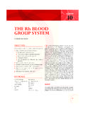
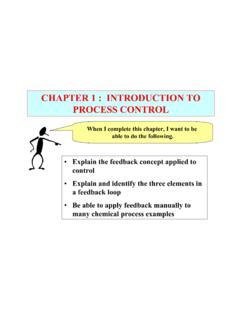
8 Figure 5-3illustrates the bones and bone markings of the leg. CPosterior superioriliac spinePosterior inferioriliac spineSacrumGreater sciatic notchIschial spineLesser sciatic notchCoccyxIschial tuberosityGreater trochanterLesser trochanterInferior ramus of pubisPubic tubercle Superior ramus of pubisIliac crestL4L3 Anterior superior iliac spineAnterior inferior iliac spineHead of femurThe coxal joint is formed betweenthe head of the femur and is a stable ball-and-socket joint thatallows movement in all acetabulum is a deep socketformed by the fusion of the ilium,ischium, and of female pelvis which isgenerally wider and more horizontallyoriented than the of femurLWBK788-Ch5_297-437_Moorecraft Edition 1 24/01/11 9:25 PM Page 299 BAAdductortubercleMedialcondyleof femurFibulaTibiaShaft of femurMediatibialcondylelGreater trochanterLesser trochanterNeck of femurAt the patellofemoral jointthe patella glides inferiorlyas the knee flexes andsuperiorly as it maintains the leverageof the surrounding musclesduring tibiofemoral joint is amodified hinge that allowsflexion, extension, andslight rotation of the kneePatellaLateral condyleof femurHead of fibulaTibal tuberosityMedialcondyleof femurShaft of femurMedialtibialcondyleGreater trochanterLesser trochanterGluteal tuberosityPectineal lineHead of femurNeck of femurThe linea aspera is a long verticalline running along the shaft of theposterior femur.
9 It is the site ofseveral muscle condyleof femurIntercondylar notchHead of fibulaLateral condyleof tibiaFIGURE 5-2 Femur, femoral bone markings, and the patella. A: Anterior view; B: Posterior view300 PART 2 INDIVIDUAL MUSCLES BY BODY REGIONLWBK788-Ch5_297-437_Moorecraft Edition 1 24/01/11 9:25 PM Page 300 Lateral condyle of the tibiaFibular headFibular shaftTibial shaftMedial condyle of the tibiaLateral malleolusCalcaneusCuboidMiddle phalanxFifth metatarsalDistal phalanxMedial malleolusTarsalsProximal phalanxFirst metatarsalDistal phalanx The proximal tibiofibular jointis a stable synovial joint allowinglittle movement. The distal tibiofibular joint is afibrous syndesmosis that allowsvery little motion, increasingstability of the low leg. The tibial tuberosity is the bump below the knee. The talocrural joint is a true hingejoint. Plantarflexion and dorsiflexionare possible here, but the distaltibiofibular joint must give slightly,allowing the talus to move posteriorlyduring endrange dorsiflexion.
10 The soleal line of the tibiamarks the attachment ofthe soleus muscle. TarsalbonesMedial malleolusLateral malleolusCalcaneusTalusCuboidFibulaNeck of fibulaABTibiaFIGURE 5-3 Bones and bone markings of the leg. A: Anterior view; B: Posterior viewCHAPTER 5 Lower Limb301 LWBK788-Ch5_297-437_Moorecraft Edition 1 24/01/11 9:25 PM Page 301 ABNeck of talusFirst cuneiform NavicularMedial longitudinal archMetatarsalsFirst proximalphalangeFirst distalphalangeMedial sesamoidDome of talusThe talus articulateswith the tibia andfibula calcaneus andtalus bear most of theweight of the tubercleof talusCalcaneusLaterallongitudinalarchPer onealtubercleTuberosityof 5thmetatarsalTransversearch PhalangesBody NeckTalusHead11234523 NavicularCuboidCuneiformsAttachment ofcalcaneofibularligamentThe metatarsals articulate withthe tarsals and the tarsal bones include the calcaneus,talus, navicular, cuneiforms, and 5-4 Bones and arches of the foot.