Transcription of SEIZURE PROPHYLAXIS IN PATIENTS WITH …
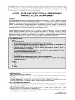
1 DISCLAIMER: These guidelines were prepared by the Department of Surgical Education, Orlando Regional Medical Center. They are intended to serve as a general statement regarding appropriate patient care practices based upon the available medical literature and clinical expertise at the time of development. They should not be considered to be accepted protocol or policy, nor are intended to replace clinical judgment or dictate care of individual PATIENTS . SEIZURE PROPHYLAXIS IN PATIENTS WITH. traumatic brain injury (TBI). SUMMARY. Post- traumatic seizures (PTS) are common complications of TBI. Early PTS are linked to a high incidence of late PTS and chronic epilepsy. CT scan is a useful modality in identifying the probability of late PTS. EEG is a promising, but currently underdeveloped technique for the prognostic evaluation of PTS. Pharmacoprophylaxis of early PTS includes phenytoin, levetiracetam, and carbamazepine.
2 Pharmacoprophylaxis of late PTS is not routinely recommended. Valproate and phenobarbital are not acceptable agents in the PROPHYLAXIS of PTS. RECOMMENDATIONS. Level 1. Phenytoin is effective in decreasing the risk of early PTS in PATIENTS with severe TBI. Valproate should not be used for early PTS PROPHYLAXIS . Phenytoin, carbamazepine or valproate are ineffective in decreasing the risk of late PTS. There are insufficient data to recommend routine PTS PROPHYLAXIS in PATIENTS with mild or moderate TBI. Level 2. Levetiracetam is an effective and safe alternative to phenytoin for early PTS PROPHYLAXIS . Routine PROPHYLAXIS of late PTS is not recommended. Level 3. Levetiracetam should not replace phenytoin as a first-line agent for PTS PROPHYLAXIS based upon efficacy and economic analysis. Carbamazepine is effective in decreasing the risk of early PTS. The following CT scan findings may indicate the need for late PTS PROPHYLAXIS (anticonvulsant therapy for longer than 7 days post injury ) (1): o Biparietal contusions (66%).
3 O Dural penetration with bone and metal fragments ( ). o Multiple intracranial operations ( ). o Multiple subcortical contusions ( ). o Subdural hematoma with evacuation ( ). o Midline shift greater than 5mm ( ). o Multiple or bilateral cortical contusions (25%). CT scan findings are superior to GCS score when used as a predictor for PTS development. EEG does not currently have a role in evaluating the need for PTS PROPHYLAXIS . EVIDENCE DEFINITIONS. Class I: Prospective randomized controlled trial. Class II: Prospective clinical study or retrospective analysis of reliable data. Includes observational, cohort, prevalence, or case control studies. Class III: Retrospective study. Includes database or registry reviews, large series of case reports, expert opinion. Technology assessment: A technology study which does not lend itself to classification in the above-mentioned format.
4 Devices are evaluated in terms of their accuracy, reliability, therapeutic potential, or cost effectiveness. LEVEL OF RECOMMENDATION DEFINITIONS. Level 1: Convincingly justifiable based on available scientific information alone. Usually based on Class I data or strong Class II. evidence if randomized testing is inappropriate. Conversely, low quality or contradictory Class I data may be insufficient to support a Level I recommendation. Level 2: Reasonably justifiable based on available scientific evidence and strongly supported by expert opinion. Usually supported by Class II data or a preponderance of Class III evidence. Level 3: Supported by available data, but scientific evidence is lacking. Generally supported by Class III data. Useful for educational purposes and in guiding future clinical research. 1 Accepted 8/27/2012. INTRODUCTION. traumatic brain injury (TBI) is a common neurologic disorder accounting for million emergency department visits and one hospitalization per 1,000 people each year in the United States (1,2).
5 Among all PATIENTS with head trauma who seek medical attention, 2% develop post- traumatic seizures (PTS) although the number varies widely depending upon injury severity. Approximately 12% of PATIENTS with severe TBI. will develop PTS, however, this risk may approach 50% when SEIZURE activity is diagnosed by electroencephalography (EEG) (3,4). Exposure to penetrating missile injuries is associated with a PTS rate as high as 50% (5-7). PTS may cause secondary brain injury as a result of increased metabolic demands, increased intracranial pressure, compromised cerebral oxygen delivery, and excess neurotransmitter release. The following are risk factors for the development of PTS (8): Alcoholism Loss of consciousness Age Focal neurologic deficits Penetrating injuries Depressed skull fracture Intracranial hemorrhage Cerebral contusions Severity of injury Retained bone and metal fragments Length of posttraumatic amnesia Lesion location Early seizures after traumatic and non- traumatic brain insults have been found to be predictive of subsequent epilepsy development (9,10).
6 While a SEIZURE during the first week after injury (early PTS) is associated with subsequent late PTS (after the first week postinjury), late PTS is correlated with an even higher rate of recurrence. Prompt and effective PROPHYLAXIS of both early and late PTS is crucial to the reduction of SEIZURE recurrence rates as well as the morbidity of recurrent seizures. When selecting appropriate medical management, it is important to differentiate between (11): Early PTS: 0 7 days after injury Late PTS: more than 7 days after injury The severity of TBI can be estimated as (3,12,13): Mild: loss of consciousness or post- traumatic amnesia less than 30 minutes or GCS 13-15. Moderate: loss of consciousness or post- traumatic amnesia for 30 minutes to 24 hours or a skull fracture or GCS 9-12. Severe: brain contusion or intracranial hematoma or a loss of consciousness or post- traumatic amnesia for more than 24 hours or GCS 3-8.
7 TBI may be assessed radiographically using the Marshall Computed Tomography (CT) Classification for Head injury (14) (Table 1). Table 1: Marshall CT Classification Category Definition Diffuse injury I - No visible intracranial pathology seen on CT. - Cisterns are present with midline shift 0-5 mm and/or lesion densities present Diffuse injury II. - No high- or mixed-density lesion >25 mL. - May include bone fragments and foreign bodies - Cisterns compressed or absent with midline shift 0-5 mm Diffuse injury III. - No high- or mixed-density lesion >25 mL. - Midline shift > 5 mm Diffuse injury IV. - No high- or mixed-density lesion >25 mL. Diffuse injury V. - Any lesion surgically evaluated (Evacuated mass lesion). Diffuse injury VI - High- or mixed-density lesion >25 mL. (Non-evacuated mass lesion) - Not surgically evacuated 2 Accepted 8/27/2012. The use of antiepileptic drugs to treat PATIENTS who have developed post- traumatic epilepsy is an accepted standard of care.
8 However, there is substantial variability among clinicians in the practice of PTS. PROPHYLAXIS . Two surveys of neurosurgeons reported that a majority prescribed antiepileptic drugs for SEIZURE PROPHYLAXIS at least some of the time, although the indications, choice of drug, and duration of treatment varied widely (15,16). Similar variability was seen in the care of head injured PATIENTS referred to a rehabilitation center (17). This review systematically analyzes available literature and proposes recommendations for SEIZURE PROPHYLAXIS in TBI PATIENTS . LITERATURE REVIEW. Imaging Modalities in PTS PROPHYLAXIS Electroencephalography (EEG). Several methods have been suggested to improve identification and monitoring of PTS onset. EEG was proposed as a potential method of prediction and early PROPHYLAXIS . Studies do not clearly associate EEG. findings with early or late PTS (18-21).
9 Vespa et al. (Class II study) employed continuous EEG monitoring to establish the incidence of convulsive and non-convulsive seizures in the ICU during the initial 14 days post- injury (22). In 52% of PATIENTS , the seizures were non-convulsive and diagnosed on the basis of EEG. studies alone. Another study by Vespa (Class II) assessed the usefulness of continuous EEG monitoring in ICU PATIENTS for determining prognosis after TBI (23). Percentage of alpha variability (PAV) was found to be a sensitive and specific method of prognosis to indicate outcomes in PATIENTS with moderate to severe TBI. within 3 days of injury . Ronne-Engstrom et al. (Class II study) established the presence of certain epileptiform activity on EEG in TBI PATIENTS , which in 67% of cases was a SEIZURE (24). Jones et al. (Class II. study) utilized 1 hour EEG monitoring for comparing phenytoin vs.
10 Levetiracetam monotherapy for PTS. PROPHYLAXIS in PATIENTS with severe TBI. They concluded that clinical seizures are difficult to identify through observation or physical examination in the early stages after severe TBI. In addition, TBI itself along with sedative and neuromuscular blockade agents used in the ICU may mask SEIZURE activity. Routine use of EEG monitoring to discern abnormal EEG patterns was encouraged. Based on the evidence outlined above, EEG appears to be an attractive option of early SEIZURE onset detection and PROPHYLAXIS , especially in the ICU setting. However, more studies are needed to precisely determine EEG patterns related to PTS. onset and establish a treatment protocol. Computed Tomography Englander et al. (Class II study) found that CT scan findings and neurosurgical procedures performed were the most useful factors in identifying moderate to severe TBI PATIENTS at highest risk for late post traumatic seizures (25).