Transcription of SKIN GRAFTS - Practical Plastic Surgery
1 SSKKIINN GGRRAAFFTTSSA skin graft involves taking a piece of skin from an uninjured area ofthe body (called the donor site) and using it to provide coverage for anopen wound. When primary closure is impossible because of softtissue loss and closure by secondary intention is contraindicated, a skingraft is the next rung on the reconstructive ladder. It is not a technicallydifficult procedure but does require some surgical skills. For a success-ful result, you need a thorough understanding of how skin GRAFTS healand how to perform the iinnffoorrmmaattiioonnAnatomy of SkinThe thickness of human skin is quite variable. The eyelids have thethinnest skin ( mm), and the thickest skin is found on the soles ofthe feet (> mm).EpidermisThe epidermis is the top portion of the skin . The outer layers of the epi-dermis are formed by essentially dead, nonreplicating cells. The inner-most layer contains the cells capable of replication, which are responsiblefor wound healing and skin below the epidermis is the dermis.
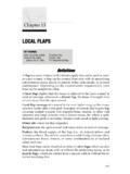
2 It is made primarily of col-lagen and is much thicker than the epidermal layer. The dermal-epidermal97 Chapter 12 KKEEYY FFIIGGUURREESS:: skin anatomy withMeshergraft thicknessSkin graftHumby knifeTying the dressing Using the dermatomein placeUsing the Humby knifeDefatting the FTSG98 Practical Plastic Surgery for Nonsurgeonsjunction is irregular and has the appearance of ridges. This anatomicarrangement accounts for the skin s strength and prevents injury fromnormal shear forces. Nerve endings, hair follicles, and sweat glands arelocated in the dermis. All skin GRAFTS must include at least a portion of thedermal layer for TissueThe subcutaneous fatty tissue below the dermis provides padding forthe skin . The base of many hair follicles and sweat glands, as well asmany important nerves for pressure sensation, reside in the subcuta-neous tissue. Because of these important skin components, I includethe subcutaneous tissue as a layer of the skin .
3 However, it is not in-cluded in a skin graft . Fat attached to the graft interferes with transportof nutrients to the important upper skin layers. Therefore, no fatshould be included in the skin graft . How a skin graft SurvivesWhen the skin graft is harvested from the donor site, it is completelyseparated from its blood supply. In its new position covering the openwound, the graft initially survives by diffusion of nutrients from thewound bed into the graft . Diffusion of nutrients keeps the skin graftCross-section of human skin showing the epidermis and dermis (derived fromtwo different germ layers). The relative thickness of skin GRAFTS is shown. Thethicker the graft , the more characteristics of normal skin it will provide. (FromCohen M (ed): Mastery of Plastic and Reconstructive Surgery . Boston, Little,Brown, 1994, with permission.) skin GRAFTS 99alive for, at most, 3 5 days. During this period, blood vessels begin togrow from the wound bed into the graft .
4 By the time the graft is nolonger able to survive by diffusion of nutrients alone, this vascular net-work has formed and becomes the primary mechanism for providingnutrients to the the first several weeks after the procedure, the skin graft looks quitered and irregular compared with normal surrounding skin . Reassurethe patient that the appearance will improve dramatically over the nextseveral months, but the skin -grafted area will never look completelynormal. It can take at least 1 year to see the final appearance of thegraft. See chapter 15, Scar Formation, for more detailsWhen is a Wound Ready for Grafting?A wound will accept a skin graft when there is no overlying deadtissue and the wound is clean, beefy red (from granulation tissue), andwithout surrounding infection. skin GRAFTS heal well over , if muscle is exposed in the wound, skin can be grafted atany time, as long as the wound is otherwise to Wound Closure with a skin GraftA wound that has exposed tendon or bone can be successfully coveredwith a skin graft only if the thin layer of tissue connecting the tendon orTTaabbllee ffoorr FFaaccttoorrss tthhaatt IInntteerrffeerree wwiitthh GGrraafftt SSuurrvviivvaall*Granulation tissue is the beefy red tissue that develops as a wound heals.
5 It has an excellent bloodsupply but also contains bacteria in its wound ( ,Debride the wound and treat it with wet-to-dry dressingssurrounding in-until the wound looks clean. Use antibiotics to clearfection, necroticsigns of surrounding infection. The skin graft can betissue over wound)done once the wound has improved in appearance andthere are no signs of surrounding in base of woundFat has a poor blood supply and may not be able to sup-port the graft . Treat the wound with wet-to-dry dressingsuntil granulation tissue* begins to appear. Then do theskin forces between Movement of the graft over the wound interferes with vas- graft and base ofcular ingrowth. The graft must be kept well secured towoundthe wound by the dressing. If the graft is on an extremity,consider using a splint for immobilization of the or serumFluid collection under the graft prevents the ingrowth ofcollection underblood vessels necessary for graft survival. Fluid collectiongraftcan be prevented by cutting holes in the graft and keep-ing the graft well secured to the wound.
6 If the graft is on the leg, the patient should be kept on bedrest, with the leg elevated at all times for at least the first 4 5 Practical Plastic Surgery for Nonsurgeonsbone (paratenon or periosteum, respectively) is intact. These connectivetissues contain the vascular structures necessary for skin graft survival. Ifthe paratenon or periosteum is absent, the graft will not survive. Underthese circumstances, some type of flap is needed for wound SSkkiinn GGrraaffttA split-thickness skin graft (STSG) is composed of the top layers of skin (the epidermis and part of the dermis). The graft is placed over an openwound to provide coverage and promote healing. The STSG donor siteis essentially a second-degree burn because only part of the dermis isincluded in the graft . The donor site will heal on its own because somedermal elements STSG is indicated in most wounds that cannot be closed primarilyand when closure by secondary intention is contraindicated.
7 It is alsoindicated for a relatively large wound (> 5 6 cm in diameter) thatwould take many weeks to heal secondarily. A skin graft providesmore stable coverage for large wounds than the scar that results fromsecondary closure. A large wound also heals more quickly with a skingraft than with dressing changes alone. The wound must be clean. Allnecrotic tissue must be removed before skin grafting, and there shouldbe no signs of infection in the surrounding of the Donor SiteBecause of the relatively large size of the graft to be taken, the patientusually requires either general or spinal anesthesia for adequate paincontrol. However, if the required graft is no more than several centime-ters in diameter, the donor site can be anesthetized by local infiltrationof tissues with lidocaine or of the Donor SiteThe most common donor site is the anterior or lateral aspect of thethigh. If the wound to be covered is on the back, try to take the graftfrom the lateral thigh, but the posterior thigh is also acceptable.
8 Use ofthe posterior thigh as a donor site is a bit more painful and difficult forthe patient to care for betadine or other antibacterial solution used to prepare the donorsite should be washed off with saline. Then the donor site should bedried. Apply a sterile lubricant ( , mineral oil, K-Y jelly) to the donorsite and to the instrument you will be using to harvest the GRAFTS 101 Procedure for Taking the GraftA thin layer of skin (epidermis with some underlying dermis) istaken with a dermatome or a Humby knife (sometimes called aWatson knife). A dermatome is powered by air or electricity, but it isnot available in all hospitals, especially in rural settings. Remember:you are nottaking full-thickness skin ; some dermis must be left at thedonor the Humby knife and dermatome have settings that can be ad-justed to set the thickness of the graft . Place the settings at ( mm). Unfortunately, these settings are often technique to ensure proper thickness of the graft is to adjustthe opening of the blade so that you can snuggly fit the beveled edge ofa no.
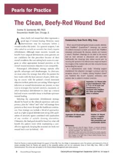
9 10 blade into the :Always check the knife settings just before you take the safety check prevents the accidental taking of too thick or too thina graft . An assistant should help to spread and flatten out the donor site byplacing tension on the skin with gauze or tongue you have a dermatome:1. Turn on the power while the dermatome is in the air before it comesinto contact with the Hold the dermatome at a 45 angle with the skin and hold it firmlyagainst the Slowly move down the donor site until you have taken the properlysized At this point do notturn off the power. Remove the dermatomefrom the skin with the power on so that the graft is completely freedfrom the donor The entire movement is evocative of landing an airplane and takingoff again right (Humby) knife. (From Padgett Instruments, Inc., with permission.)102 Practical Plastic Surgery for NonsurgeonsIf you have a Humby knife:1. Hold it with the sharp edge at about a 45 angle with the With a back-and-forth motion run the knife over the tight When you have taken a large enough graft , continue the back-and-forth motion, and twist your wrist into supination to remove theknife from the skin .
10 Another option is to stop the knife movementand then use a scalpel to cut the skin graft from the donor site at theHarvesting a split-thickness skin graft with a power-driven dermatome. (FromCohen M (ed): Mastery of Plastic and Reconstructive Surgery . Boston, Little,Brown, 1994, with permission.)Harvesting a split-thickness graft with the Humby knife. (From McCarthy JG(ed): Plastic Surgery . Philadelphia, Saunders, 1990, with permission.) skin GRAFTS 103blade edge. You may need to open the knife fully to remove the skinfrom the of the skin GraftIt is best to cut multiple slices in the graft to prevent blood and serumfrom accumulating under the graft . The cuts also help to expand thegraft, allowing you to take a graft that is slightly smaller than the openwound. Use the tip of a knife or a small scissors to create the cuts in thegraft. Some operating rooms have special equipment, called meshers,for this purpose. The mesher is a hand-cranked instrument that createspie-cuts in the , The mesher, a device used tomake fine cuts in skin GRAFTS .