Transcription of SOFAMOR DANEK ZEPHIR - MT Ortho
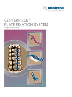
1 SOFAMOR DANEKas described by:Richard Assaker, University HospitalLille, FrancePaul McCormick, Presbyterian HospitalNew York, New YorkZEPHIR Anterior Cervical Plate SystemSurgical TechniqueDear Fellow Colleague:Anterior cervical plating has become widely accepted when anterior spinal fusion surgery isperformed. Through our surgical experience over the past years, we have found the need for a simple to use, low profile plate, which maintains the existing standards of strength for anteriorcervical ZEPHIR Anterior Cervical Plate System, developed in Lille, France, represents an advance incervical plate fixation. For the surgeon, the ZEPHIR System s minimized height and narrowtransverse width enhance visualization and plate manipulation for precise plate placement whilereducing the amount of traction on the trachea and esophagus. The rotating drill guides,self-tapping screws and integrated antimigration cap simplify plate fixation and reduce operativetime.
2 Finally, the flexibility of the ZEPHIR System through variable angulation of screw trajectory,multiple screw lengths and revision screws allows the surgeon to individually tailor theconstruct to each patient s anatomy. We believe that these design features make implanting theZEPHIR System plate a nearly seamless component of the anterior cervical decompression andstabilization ZEPHIR System has been tested following the ASTM testing standards and was found toperform equal to or better than other clinically available systems. Prior to its launch, the ZEPHIRS ystem was used worldwide by a group of surgeons to enhance the instrumentation and theimplants and to measure its clinical following monograph describes the ZEPHIR System as well as some of our personal thoughtsreflecting our current operative ,Richard Assaker, McCormick, University HospitalColumbia Presbyterian HospitalLille, FranceNew York, New YorkINTRODUCTIONFIGURE12 PATIENT POSITIONING/ANTERIOR APPROACHThe patient is placed in the supine position with the head in slight extension.
3 The posteriorcervical spine is supported to establish and maintain normal cervical lordosis. The surgeon mustthen choose a right- or left-sided approach to the cervical exposing the cervical spine, the self-retaining retractor is placed to provide optimalvisualization (Figure 1). A vertebral body distractor may be used. The distraction pins arepositioned midline in the vertebral bodies adjacent to the level to be treated. The distractor isplaced over the pins and the appropriate amount of distraction is forceps, curettes, and kerrisons may be used to remove the disc material and cartilage toexpose the posterior longitudinal ligament. Bone graft is then positioned between the For C2 fixation it is always necessary to contourthe superior aspect of the plate. I bend the platejust caudally with respect to the antimigration cap. R. Assaker, STEP13 PLATE SELECTION & CONTOURINGA nterior osteophytes are removed from the exposed vertebrae so that the plate may sit flush/evenlyon the anterior cortex.
4 The plate length should be defined according to the chosen screw lengthand angulation so that it does not interfere with the adjacent unfused disc plate is machined into a lordotic curve. However, the Plate Bender can be used to increase or decrease lordosis as necessary (Figures 2 and 3). Bending at the extremities of the plate is notrecommended as it could damage the antimigration cap Selection Plate ContouringFIGURE4 FIGURE6 FIGURE7 STEP24 PLATE POSITIONING Alternatively, a screw distractionpost can be utilized to buttresseither end of the plate. P. McCormick, pick up a plate with the Plate Holder, push the button on the proximal end of the Plate Holderand insert the tip into any screw hole or central slot in the plate. Release the button to engage theplate (Figure 4).Prefixation Pins can be used to assist positioning and to stabilize the plate in the midline prior toscrew insertion. To open the jaws of the Prefixation Pin Holder, pull the flange on the shaft towardthe handle.
5 Place it over a Prefixation Pin in the sterilization case and release the flange to engagethe pin. Insert the pin through one of the four small holes along the midline of the plate (Figure 5).Do not insert the pin through the bone screw holes. Slide the jaws of the instrument to the sideand then lift it up to clear the pin. Next, pick up a second Prefixation Pin from the sterilizationcase and insert it into one of the small holes at the other end of the alternative use for the Prefixation Pins is to assist in placing the plate along the midline. First,place the superior pin in the midline of the superior body (Figure 6). Insert the needle portion ofthe pin only. Next, place the notch in the end of the plate against the bulb portion of the the plate is precisely positioned, insert the inferior pin through the plate in one of the smallmidline holes at the other end of the plate (Figure 7). Plate prefixation allows me toprecisely check the positioning andthe length selection of the plate.
6 R. Asssaker, Prefixation Pins hereInsert Prefixation Pins hereOption 1: Mono Drilling GuideFirst, position the Mono Drilling Guide intothe bone screw hole in the plate. Then apply slight downward pressure until theleg support of the Drilling Guide comes intocontact with the plate (Figure 8).The Mono Drilling Guide is designed toprovide a maximum of 16 of angulationduring the drilling step. To achieve lessangulation, pivot the guide toward the end of the plate. The range of cephalad/caudalangulation is from 0 to 16 (Figures 8a and 8b).Loosen the nut with the Universal Handle andinsert the tri-flat drill bit into the the nut to securely hold the drill bit. If using power to drill, the tri-flat or thecircular tip drill bit can be used. The drillingdepth is fixed to 13mm, (Figure 8b) with off-center drill hole for graftcompression (Figure 8c), which will beachieved as the bone screw is handle of the Mono Drilling Guiderotates for optimal handle The Mono Drilling Guide is mostfrequently used.
7 First, I drill twodiagonally opposite screw holes andthen I partially insert the screws. R. Assaker, The 180 rotation of the drill guideenhances the stability and control ofdrilling by allowing me to comfortablydrill both superior and inferior holeswith my dominant hand. P. McCormick, 0 I prefer to angle the rostral screws 5 to 10 superiorly to optimize load sharing andpermit some graft settling allowed by thissemi-constrained system. I place the caudal screws perpendicular to the plate ( 0 angulation). P. McCormick, OFDRILLED HOLECENTER OFBONE SCREWHOLEO ption 2: Dual Drilling GuideThe angulation is first determined by adjusting the leg support of the Dual Drilling Guide (Figure 9).Two marks are engraved on the leg support. If the leg support s upper mark is set to the top of theplatform, the drilling angulation is 0 (Figure 9a). The lower mark yields a 16 drilling angulation(Figure 9b).
8 Angulation of the Dual Drilling Guide should not exceed 16 .Place the double barrel of the Dual Drilling Guide into the bone screw holes of the plate and slightlylower the guide until the leg support sits on the both holes successively through the DrillingGuide. The drilling depth is fixed to 13mm, with an offset of for graft compression. (See Figure 8c,Page 5)Once the drilling is performed, remove the Drilling Guide. The handle of the Dual DrillingGuide rotates for optimal handle 0 FIGURE9aFIGURE9bSTEP36 The construct could be constrained by aimingthe screw hole at 0 . For a hybrid construct,one set of screws could be directed at 0 andthe other set between 0 and 16 . R. Assaker, MarkPlatformLower MarkPlatformDRILLINGFIGURE9 The standard bone screw diameter is the revision/central slot bone screwdiameter is These bone screws areself-tapping and come in 11, 13, 15, and 17mmlengths. The Screwdriver consists oftwo pieces, the Sleeve and the the Sleeve from the Shaft and use the Sleeve to pick up the appropriate diameterand length bone screw (Figure 10).
9 Then placethe Shaft through the center of the Sleeve toengage the cruciate drive in the screw (Figure 11).FIGURE10 STEP47 FIGURE11 BONE SCREW SELECTION To increase efficiency of theplate insertion, the scrub nurseloads the screws while thesurgeon drills the screw surgeon s eyes do not leavethe operative field at any timeduring plate insertion. P. McCormick, insert two bone screws diagonallypositioned in the plate (Figure 12). Removethe Prefixation Pins with the Pin Holder. Fullyinsert the two remaining screws (Figure 13).Complete final tightening of the first two compression of the graft is achieved by thecombination of the offset drilling ( ) andthe screw head interference with the platescrew holes (Figure 14). The red dot in theillustration represents the drilled hole; the blackdot represents the center of the bone screwhole. The drilled holes are off center by hole. As the screw heads are seated in theplate, they pull the vertebral bodies towardeach other, resulting in of graftcompression on each end of the plate, of total performing a multi-level procedure andintermediate vertebral body or graft fixation isdesired, the diameter screws must beused in the central slot (Figure 15).
10 Thesescrews are designed to self-lock in the centralslots via an interference between the minordiameter of the screw and the sides of the slot. It is recommended that the pilot hole be drilledperpendicular to the Remove Prefixation Pins prior to finaltightening of the bone screws tomaximize the compression capabilities. R. Assaker, OF DRILLED HOLECENTER OF BONE SCREW HOLEBONE SCREW INSERTION Retraction of the Screwdriver Sleeveonce the screw is firmly engaged inthe bone improves visualization andfacilitates final screw tightening. P. McCormick, Antimigration Cap Pusher is assembledonto the Universal Handle by inserting the UNLOCK tip into the handle and tighteningthe nut on the translation of both antimigration caps isobtained by inserting the LOCK tip into the gap between the plate and the cap and giving the instrument a quarter turn (Figure 16). Whenproperly advanced, the tips of the antimigrationcaps will partially cover the bone screw heads,thus preventing screw migration.