Transcription of Structure and Mechanism of Alkaline Phosphatase
1 Annu. Rev. Biophys. Biomol. Struct. :441~3 Copyright ~ 1992 by Annual Re~iews Inc. All rights reservedSTRUCTURE AND MECHANISMOF Alkaline PHOSPHATASEJ oseph E. ColemanDepartment of Molecular Biophysics and Biochemistry, Yale University,New Haven, Connecticut 06510 KEY WORDS~zinc enzymes, enzyme mechanisms ( Alkaline Phosphatase ), crystalstructure ( Alkaline Phosphatase ), 31p NMR ( Alkaline phospha-tase), 1~3Cd NMR ( Alkaline Phosphatase )CONTENTSINTRODUCTION ..442 GENERAL Structure OF E. COLI Alkaline Phosphatase ..443 SUMMARY OF SUBSTRATE SPECIFICITY AND KINETICS OF Alkaline Phosphatase ..443 THE METALLOENZYME NATURE OF Alkaline Phosphatase ..453 COORDINATION CHEMISTRY AT THE ACTIVE CENTER OF Alkaline Phosphatase ..456Zn l ( A ) Coordination ..460Zn2(B) Coordination ..460M#3(C) Coordination ..460 Enzyme-Bound Phosphate in the E" P Intermediate ..461 Enzyme-Bound Phosphate in the E-P Intermediate.
2 461 CHANGES IN ACTIVITY OF Alkaline Phosphatase ON SUBSTITUTING CADMIUM, COBALT,OR MANGANESE FOR THE NATIVE ZINC ION ..463 CONCLUSIONS ON Structure AND Mechanism OF Alkaline Phosphatase DERIVEDFROM MULTINUCLEAR NMR ..464 CORRELATION OF Structure AND Mechanism ..468 Michaelis Complex with a Phosphate Monoester ..468 The Phoshoseryl Intermediate ..469 Hydrolysis of the Phosphoseryl Intermediate ..469 Dissociation of the Product, Inorganic Phosphate, the Rate-Limiting Step ..470 The Phosphotransferase Reaction ..471 SUMMARY OF THE Mechanism OF Alkaline Phosphatase AS BASED ON SOLUTION DATAAND THE CRYSTAL Structure ..473 SITE-D~RECTED MUTANTS OF e. COLZ Alkaline Phosphatase ..476 Serl02 ~ Cysl02, .4la102, Leul02 ..477 Arg166 ~ Lys166, Glu166, Ser166, ,4la166 ..477 Lys328 ~ His328, Ala328 ..478 SUMMARY ..4804411056-8700/92/0610-0441 $ Rev. Biophys. Biomol.
3 Struct. :441-483. Downloaded from University of Wisconsin - Eau Claire (McIntyre Library) on 11/10/06. For personal use COLEMANINTRODUCTIONA lkaline Phosphatase is often cited as the most frequently referencedenzyme (55). This fact relates more to the widespread use of alkalinephosphatase activity in human serum as an enzymatic signal for a varietyof disease states involving particularly the liver and bone, than to a greaternumber of investigations directed at the molecular properties of theenzyme. The emergence in the literature of the enzyme Alkaline phos-phatase began around 1907 when Suzuki et al first suggested that phos-phatases constituted a separate class of eukaryotic enzymes (70). By 1912,the enzyme we now know as Alkaline Phosphatase was defined by the workof Grosser & Husler (40) and von Euler (75), who showed that while was present in a variety of tissues, the enzyme, which could hydrolyzeglycerophosphate and fructose 1--6 diphosphate, was present in highestamount in intestinal mucosa, von Euler & Funke (76) used the wordphosphatase for the first time in 1912.
4 The enzyme from intestinal mucosa,particularly calf intestine, became the prototype for investigators exploringthe properties of the enzyme until 1961 did Engstrom discover that the intestinal enzyme formeda phosphoseryl residue when incubated with phosphate esters at low pH(27, 28). The enzyme s catalysis of 180 exchange into inorganic phosphatestrongly supported the notion that the phosphoserine is a significant inter-mediate on the catalytic pathway (3, 65, 69). The demonstration thattransfer of phosphate by the enzyme from an ester to a second alcoholleads to retention rather than inversion of configuration around the phos-phorous (46) supported the conclusion derived from much previous evi-dence that the Mechanism involves two sequential in-line nucleophilicattacks at phosphorous, the first by the hydroxyl of Ser 102 on the incomingphosphomonoester and the second on the phosphoseryl intermediate bysolvent water or an alcohol acceptor (32-34, 38).
5 In the late 1950s and early 1960s, investigators discovered that Esch-erichia coli possessed an Alkaline Phosphatase that was derepressible byphosphate starvation (21, 31, 42, 72). Its gene, phoA, was part of the phoregulon consisting of the group of genes in E. coli whose products areinvolved in phosphate transport and metabolism (4, 22, 38, 53) (for bib-liography, see 22, 38). The E. coli enzyme had similar catalytic properties,similar pH-rate profile, and formed the same phosphoseryl intermediate asthe intestinal enzyme (66, 67). The amino acid sequences of the mammalianenzymes, derived from their cDNA sequences, can be fit into the primarystructure of the bacterial enzyme, with the proper adjustments for someinsertions and deletions (47). With such adjustments, most of the criticalactive-site residues described below are conserved between the eukaryoticand bacterial Rev.
6 Biophys. Biomol. Struct. :441-483. Downloaded from University of Wisconsin - Eau Claire (McIntyre Library) on 11/10/06. For personal use PHOSPHATASE443In 1962, the E. coli Alkaline Phosphatase joined the class of zinc metallo-enzymes with the demonstration by Plocke et al (62) that the enzymecontained stoichiometric amounts of zinc, a finding also confirmed in laterstudies of the calf intestinal enzyme (30). These early studies showed thatZn(II) is required for activity, and many subsequent studies have confirmedthis observation, including the demonstration that the metal is required forinitial phosphate binding and thus for the formation of the phosphoserylintermediate (3). The great stability over a wide range of pH of the metal-free apophosphoryl enzyme, which can be formed by removal of the metalion from the Cd(II) enzyme, demonstrates that the metal ion is requiredfor dephosphorylation of the phosphoseryl intermediate as well (18).
7 The most detailed information on the Structure and function of theenzyme is available for the E. coli enzyme. This review is a synthesis ofthe solution data and the recently completed crystal structures of the Alkaline Phosphatase and its two phosphoenzyme intermediates at ~ and resolution, respectively (48). The phosphoenzymes are E the noncovalent complex formed between inorganic phosphate and theenzyme, and E-P, the covalent or phosphoseryl intermediate, formed bythe phosphorylation of Structure OF E. COLI ALKALINEPHOSPHATASEA lkaline Phosphatase exists in the periplasmic space of E. coli as a dimerof identical subunits each containing 429 amino acids (11). The fourCys residues are present as two intrachain disulfides. The monomers aresynthesized as a preenzyme containing a Leu-rich signal peptide of 22residues (7, 45, 56, 57). Processing occurs via a signal peptidase aftersecretion through the membrane (17).
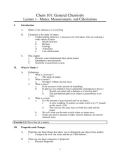
8 Recent data suggest that formationof active enzyme upon dimerization may be a complex process involvingsome modulator molecules (J. F. Chlebowski, personal communication).A comparison of the amino acid sequences of the phosphorylated peptidesisolated from the enzyme with the complete amino acid sequence showedthat the phosphorylated residue was Serl02. Figure la shows the overallshape and polypeptide conformation found in the crystal Structure of thedimer. Figure lb shows a ribbon diagram of the secondary Structure ofthe OF SUBSTRATE SPECIFICITY ANDKINETICS OF Alkaline PHOSPHATASEA lkaline Phosphatase is thought to be strictly a phosphomonoesterase,although one recent investigation found a low phosphodiesterase activityAnnual Rev. Biophys. Biomol. Struct. :441-483. Downloaded from University of Wisconsin - Eau Claire (McIntyre Library) on 11/10/06.
9 For personal use 1 [a) Alpha-carbon trace of the dimer of E . coli Alkaline Phosphatase . The non- crystallographic two-fold axis is vertical and the maximum dimension of the dimer is hori- zontal. The three metal ions at each active center are shown as spheres and the two active sites of the dimer are 30 A apart. ( b ) Ribbon drawing of the monomer of E. coli Alkaline Phosphatase . The three metal ions are shown as stippled spheres, Znl, Zn2, and Mg as indicated. The center of the monomer consists of a 10-stranded 8-sheet flanked by 15 helices of varying lengths. A second 3-stranded 8-sheet and an a-helix form the top of the molecule in this view. Reprinted from Ref. 48. Annual Rev. Biophys. Biomol. Struct. :441-483. Downloaded from University of Wisconsin - Eau Claire (McIntyre Library) on 11/10/06. For personal use Phosphatase 445(J. F. Chlebowski, personal communication).]
10 The enzyme hydrolyzes notonly oxyphosphate monoesters (23, 29, 41, 63), but also a variety of (58, 59) and S-phosphorothioates (19), phosphoramidates (63), thiophos-phate, and phosphate (3, 19, 65, 69); hydrolysis of the latter group reflected by the catalysis of the exchange of 180 from H2180 into inorganicphosphate (3, 65, 69) or the release of H2S from thiophosphate (19). enzyme has an Alkaline pH maximum, and the rate follows an approxi-mately sigmoid pH-rate profile with an apparent pK, of ~ (19, 34, 50,63). Table 1 summarizes substrate Structure , reaction products, and k~tvalues. In the case of oxyphosphate monoesters, the koat is apparentlyindependent of the R group, which can vary from a large protein moleculeto a methyl group (63). This finding reflects the fact that either the de-phosphorylation of E-P or the dissociation of the product, Pi, is the rate-controlling step depending on pH.