Transcription of Teaching Case of the Month - Respiratory Care
1 Joshua O Benditt MD, Section EditorTeaching case of the MonthBullous Lung Disease or Bullous Emphysema?Ritesh Agarwal and Ashutosh N AggarwalIntroductionBullous lung disease is an entity characterized by thepresence of bullae in one or both the lung fields, withnormal intervening ,2On the other hand, bullousemphysema is the presence of bullae in a patient withchronicobstructivepulmonarydisease(C OPD),andischar-acterized by the presence of centrilobular emphysema inthe nonbullous 5To select patients who are morelikely to benefit from bullectomy (bullous lung disease), aproper preoperative assessment is tomography (CT) can locate the bullae withconsiderable accuracy, even when their presence is notsuspected on the basis of clinical and radiographic data. Italso helps in assessing the extent and localization of bullaeand associated diffuse nonbullous emphysema.
2 Pulmonaryfunction tests (PFTs) are also important tools in the as-sessment of these conditions. In addition to helping withthe diagnosis, PFTs can help make an objective assess-ment of the severity of the underlying disease and monitorthe response to treatment. The case we describe belowillustrates the preoperative evaluation of a patient withgiant bullous lung disease and the role of CT and PFT inthe management of a patient presenting with giant ReportA 50-year-old male nonsmoker presented with historyof progressive exertional dyspnea of 2 years duration. Atpresentation to our institute the patient had Medical Re-search Council grade III dyspnea. There was no history ofcough, chest pain, or hemoptysis. General physical exam-ination was normal. Examination of the Respiratory systemrevealed absent breath sounds in the whole of the righthemithorax.
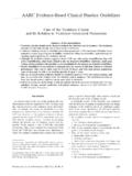
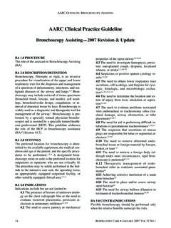
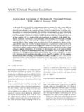
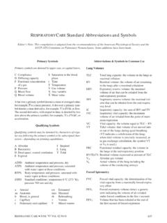
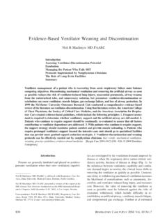
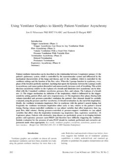
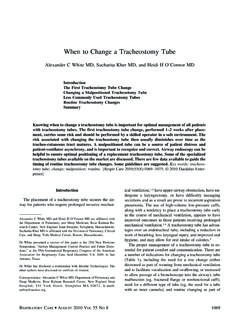
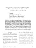
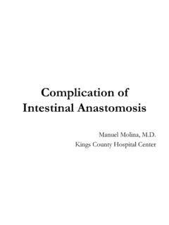
3 A chest radiograph showed multiple air-filledspaces in both the lungs, with the largest in the right hemi-thorax, occupying the whole of the right lung (Fig. 1). CTchest scan confirmed this finding, and also showed ab-sence of emphysema in the nonbullous lung (Fig. 2). Aprovisional diagnosis of bullous lung disease was showed a restrictive defect and confirmed the pres-ence of noncommunicating air spaces, as evidenced by difference between the total-lung-capacity (TLC)value measured via body plethysmography and the TLCvalue measured via the helium-dilution technique (Table1). In view of his symptoms, the patient underwent bul-lectomy. The postoperative chest radiograph showed com-plete expansion of the right lung (Fig. 3). PFTs repeated 3months postoperatively showed a difference of L be-tween the TLC measured via body plethysmography ver-sus via the helium-dilution technique; that difference wasdue to small bullae in the left lung (Table 2).
4 A familyscreening to look for similar abnormalities was patient has been asymptomatic on regular follow-upfor the last 2 Agarwal and Ashutosh N Aggarwal are affiliated with the De-partment of Pulmonary Medicine, Postgraduate Institute of Medical Ed-ucation and Research, Chandigarh, : Ritesh Agarwal, Department of Pulmonary Medicine,Postgraduate Institute of Medical Education and Research, Sector 12,Chandigarh 160012, India. E-mail: 1. Chest radiograph showing a large bulla in the right MAY2006 VOL51 NO5 DiscussionBullous lung disease is different from bullous emphy-sema in that bullous lung disease has bullae with structur-ally normal intervening lung, whereas bullous emphysemahas bullae associated with more diffusely abnormal lungparenchyma because of COPD. Giant bullous lung dis-ease, as seen in our patient, is said to be present if thebullae occupy at least one third of the hemithorax andcompress the surrounding lung ,either via videothoracoscopy or conventional thoracotomy,is the treatment of choice for giant bullous lung disease,even if is indicated for symp-tomatic patients who have incapacitating dyspnea or chestpain, and who have complications related to bullous dis-ease such as infection or needs to be differentiated from lung-vol-ume reduction surgery (LVRS), which is surgical removalof 20 30% of nonbullous emphysematous lung from eachside.
5 The recently published National Emphysema Treat-ment Trial7showed that LVRS benefits selected subgroupsof COPD patients who have upper-lobe disease and poorexercise capacity. Specifically, LVRS improves 6-min-walk distance, forced expiratory volume in the first second(FEV1), dyspnea score, and quality-of-life score, and de-creases residual volume and the need for supplementaloxygen. However, patients with FEV1 20% of predictedand either homogenous emphysema or carbon-monoxide-diffusion capacity 20% of predicted do not benefit fromFig. 3. Postoperative chest radiograph. The right lung is fully 1. Preoperative Pulmonary Function Test ResultsVariable PredictedMeasured viaHelium DilutionMeasured via BodyPlethysmographyObserved%ofPredictedO bserved%ofPredictedFVC (L) (L) (% ofpredicted) (L) (L) forced vital capacityFEV1 forced expiratory volume in the first secondTLC total lung capacityRV residual volumeTable 2.
6 Postoperative Pulmonary Function Test ResultsVariablePredictedMeasured viaHelium DilutionMeasured via BodyPlethysmographyObserved%ofPredictedO bserved%ofPredictedFVC (L) (L) (% ofpredicted) (L) (L) forced vital capacityFEV1 forced expiratory volume in the first secondTLC total lung capacityRV residual volumeFig. 2. Computed tomography chest scan confirming the presenceof a large bulla in the right hemithorax. There is no centrilobularemphysema in the contralateral ORBULLOUSEMPHYSEMA?RESPIRATORYCARE MAY2006 VOL51 NO5533 LVRS and have unacceptable perioperative , taking a corollary from the National EmphysemaTreatment Trial, some patients with bullous emphysemamay also benefit from bullectomy. However, LVRS hasdistinct indications applicable only to a subset of patients,and with different expectations and outcomes than selection remains one of the most important as-pects of successful surgery, since bullous lung disease isassociated with excellent postoperative outcomes, whereassurgery for bullous emphysema is not very rewarding,except probably in a select group of ,8In general,the freedom from long-term return of dyspnea is propor-tional to the quality of the remaining lung after CT is an important tool for preoperativeassessment, because it can identify underlying centrilobu-lar emphysema, which is synonymous with a diagnosis ofbullous emphysema.
7 Moreover, high-resolution CT alsoallows assessment of associated diseases such as bronchi-ectasis, infected cysts, pleural disease, and pulmonary ,5 PFTs can also differentiate between the 2entities. PFT values from a patient with bullous lung dis-ease typically show a restrictive defect, whereas those froma patient with bullous emphysema show an obstructivedefect. In addition to helping in differential diagnosis, PFTsalso help in quantifying the size of bulla and objectivelydocumenting postoperative is a difference between the lung volume mea-sured via the helium-dilution technique and that measuredvia body plethysmography. In the former, the subject rap-idly breathes in and out of a reservoir that contains aknown volume of gas and a trace amount of helium (aninert gas, very little of which absorbs into the pulmonarycirculation).
8 The helium is diluted by the gas that waspreviously present in the lung. With the knowledge of thegas in the reservoir and the initial and final helium con-centrations, the functional residual capacity and the TLCcan be calculated. The helium-dilution technique may un-derestimate the exact volume of gas in the lung because ofinadequate time to equilibrate with slowly communicatingand noncommunicating air spaces such as bullae. How-ever, lung volume can be more accurately measured andshould be measured in these cases, with body plethysmog-raphy, which measures the total volume of the thorax. Infact, the difference in TLC between the 2 techniques (bodyplethysmography minus helium-dilution) approximates thevolume of the bullae. In our case , there was substantialclinical improvement with decrease in bulla volume, doc-umented on postoperative PFT (bulla volume fell L to L).
9 In conclusion, this case exemplifies the importance ofselecting the correct pulmonary function test (body pleth-ysmography) for measuring lung volume, and the utility ofCT in the evaluation of patients with bullous lung presence of bullae and the etiology (in this case , bul-lous lung disease) was confirmed by high-resolution suggested the etiology and confirmed physiologicimprovement after Roberts L, Putman CE, Chen JTT, Goodman LR, Ravin CE. Van-ishing lung syndrome: upper lobe bullous pneumopathy. Rev In-teram Radiol 1987;12:249 Murphy DM, Fishman AP. Bullous diseases of the lungs. In: Fish-man AP, Elias JA, Kaiser LR, Fishman JA, Senior RM, Grippi MA,editors. Fishman s pulmonary diseases and disorders. New York:McGraw-Hill; 1998:849 Stern EJ, Webb WR, Weinacker A, Muller NL. Idiopathic giantbullous emphysema (vanishing lung syndrome): imaging findings innine patients.
10 AJR Am J Roentgenol 1994;162(2):279 Morgan MD, Strickland B. Computed tomography in the assessmentof bullous lung disease. Br J Dis Chest 1984;78(1):10 Carr DH, Pride NB. Computed tomography in pre-operative assess-ment of bullous emphysema. Clin Radiol 1984;35(1):43 Greenberg JA, Singhal S, Kaiser LR. Giant bullous lung disease:evaluation, selection, techniques, and outcomes. Chest Surg Clin NAm 2003;13(4):631 Fishman A, Martinez F, Naunheim K, Piantadosi S, Wise R, Ries A,et al; National Emphysema Treatment Trial Research Group. A ran-domized trial comparing lung-volume-reduction surgery with med-ical therapy for severe emphysema. N Engl J Med 2003;348(21):2059 Fitzgerald MX, Keelan PJ, Cugell DW, Gaensler EA. Long-termresults of surgery for bullous emphysema. J Thorac Cardiovasc Surg1974;68(4):566 ORBULLOUSEMPHYSEMA?