Transcription of The ECHO Suggests Pulmonary Hypertension. What Next?
1 The echo Suggests Pulmonary hypertension . what Next? Dr. Jonathon Langridge, , MD, FRCPCC ardiovascular Respiratory Conference, 2016I have no conflicts of interest to declare1) Review definition and classification of PH2) Discuss presentation and diagnostic approach3) Overview of treatment strategies (Recently published: 2015 ESC/ERS Guidelines)Definition PH refers to elevated Pulmonary arterial pressure Pressure elevation in Pulmonary arterial systemalone (increased resistance or flow) OR2 to pressure elevation in Pulmonary venoussystem PH defined as mPAP 25 mmHg at rest Upper limit of normal mPAP20 mmHg mPAP21-24 mmHg considered borderline :-Unclear clinical significance-May progress to significant disease(especially idiopathic, family hx, CTD)-Warrants monitoring Thin muscular wall chamber Designed to deliver venous return to a low pressure/low resistance Pulmonary circulation Limited contractile reserve to compensate for increased PVR Chronic rise managed in part by RVH RV dilatation to increase preload Ultimately RV Categorizes multiple clinical conditions into fivegroupsbased on pathophysiology, natural hxand response to treatment Previous.
2 Primary PH (no identifiable cause)Secondary PH (identifiable cause) Nowrecognized that some types of secondary PH have similar pathophysiology and response to Rxas primary PH ( familial, CTD, portal HTN,HIV, drug-induced, congenital heart disease) Pulmonary arterial hypertension (Group 1)-Diseases with 1 vasculopathyof small PAs-Absenceof significant:Left heart disease (Group 2)Lung disease and/or hypoxemia (Group 3)Thromboembolic disease (Group 4)Rare diseases/unclear mechanism (Group 5)-Includes: IPAH, heritable PAH and associatedconditions (CTD, portal HTN, HIV, drugs, CHD) Group 1: Pulmonary arterial hypertension (PAH) Group 2: PH owing to left heart disease Group 3: PH owing to lung disease and/or hypoxemia Group 4: Chronic thromboembolic PH (CTEPH) Group 5: PH with unclear/multifactorial mechanismsWHO ClassificationPresentation Symptoms of PH nonspecific often leads to delayin diagnosis Mainly related to progressive RV dysfunction Initial symptoms typically induced by exertion Progressive exertional dyspneamost common Chest pain, syncope, fatigue With overt RV failure abdominal distension, ankle edema Less common symptoms due to mechanicalcomplications: hemoptysis, wheeze, hoarseness Symptoms of associated conditions Often normal in early stages Classic signs as RV hypertrophy and failure develop Signs.
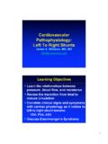
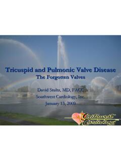
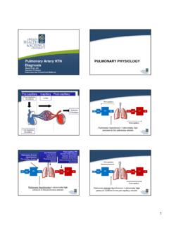
3 Left parasternal liftloud P2, right ventricular S3pansystolicmurmur of TRelevated JVPpulsatile hepatomegalyascites, peripheral edema Also findings of associated conditions Can provide supportive evidence of PH Normal EKG does not exclude PH and abnormaldoes not confirm the diagnosis Abnormal EKG more likely in severe PH Abnormalities:Right axis deviationRight atrial enlargementRV hypertrophy/strain patternRight bundle branch blockPulmonary hypertension : EKGRAD, Right atrial enlargement, RV hypertrophy, RV strainPulmonary hypertension : EKGRAD, Right atrial enlargement, Right bundle branch block Normal CXR does not exclude the diagnosis Classic abnormalities:-central Pulmonary artery dilatation-attenuation of peripheral vessels ( pruning )-RA/RV enlargement-findings of underlying etiology (eg.)
4 ILD,COPD, chest wall deformity, LV failure) Pulmonary hypertension : CXRP rominent main PA, dilated right interlobarPAPulmonary hypertension : CXRP rominent main PA, RA enlargement, peripheral pruningPulmonary hypertension : CXRR ight ventricular enlargement Classic finding isolated reduction in DLCO May see mild restrictive defect ABGs: hypoxemia, chronic respiratory alkalosis Otherwise findings of significant lung disease(WHO Group 3) Non-invasive screening tool for PH Utilities:1) Estimate Pulmonary artery systolic pressure2) Assess RV thickness, RA/RV size, RV function3) Identify 2 causes: LV systolic/diastolic dysfxn,left sided valvulardisease, intracardiacshunt Pulmonary artery systolic pressure (PASP) or RVSP estimated based on dopplerassessment of peaktricuspid regurgitantjet velocity (TRV) Calculation taking into account right atrial pressure(RAP) with simplified Bernoulli equation:PASP = (4xTRV2) + RAP Limitations:1) Cannot measure without significant tricuspid regurg2) TRV technically difficult to measure3) Can under/overestimate true PA systolic pressure Usual PASP (or RVSP) cut-off = 40 mmHg(correlates with mPAP25 mmHg) Better:-PASP > 50, associated PH findings: PH likely-PASP < 36, no ass dPH findings: PH unlikely Associated findings of PH ( the company it keeps ).
5 1) RVH (wall thickness > 5 mm)2) RV dilatation (RV/LV> , septal flattening)3) RA enlargement4) RV systolic dysfunction (TAPSE<15 mm)5) Functional TR severityPulmonary hypertension : echo Gold standard for diagnosis of PH (mPAP 25) Rule out PH due to left heart disease (Group 2):-PCWP 15 mmHg-TPG 12 mmHg, PVR < 3 WU Versus Group 1 (PAH) and Groups 3-5-PCWP < 15 mmHg-TPG > 12 mmHg, PVR > 3 WU Low threshold for left heart catheterization with direct measurement of LVEDPD iagnostic Approach Most common cause of Pulmonary hypertension *Most common cause of RHD is LHD* Elevated mPAPoccurs as a result of elevatedleft heart filling pressures Important to recognize because:1) Presence of PH in patients with LHD associated withreduced survival2) Dictates proper management= optimal treatment ofleft heart disease Etiologies:1) Heart failure with reduced EF (HFrEF)-`Systolic Dysfunction`-Ischemic vs.
6 Nonischemiccardiomyopathy2) Heart failure with preserved EF (HFpEF)-`Diastolic Dysfunction`-Most common cause of PH-LHD, often neglected-Difficult to distinguish from PAH3) Left sided valvulardisease (mitral, aortic)4) Other: Constrictive pericarditis, Restrictive CM Clues:-older age (>60)-orthopnea, PND-atrial fibrillation (uncommon in PAH)-hxof CAD, HTN, DM-left sided S3, S4, murmurs- echo : EF<50%, LAE, LVHD iastolic dysfunction (insensitive)Mod-Severe AV, MV dysfunction1) Isolated post-capillary PH Early in PH-LHD Elevated mPAPonly due to elevated LH filling pressures Severity of PH proportional to left sided pressures Pulmonary venous HTN , Passive PH Normal PVR (< 3 WU), normal TPG ( 12 mmHg) mPAPnormalizes with reduction in LH pressures to normal2)Combined Post/Pre-capillary PH Chronic elevation in LH filling pressures leading to vasculopathyof small Pulmonary arteries Vasoconstriction, endothelial dysfunction, remodeling Elevated mPAPdisproportionate to left sided pressures Mixed PH , Out-of-proportion PH Elevated PVR (> 3 WU), elevated TPG (> 12 mmHg)
7 MPAPdoesn t normalize with normalization of LH pressures ? Role for targeted PAH therapy1) Chronic Lung Disease PH is a common complication Most commonly COPD, ILD, and combined pulmonaryfibrosis and emphysema (CPFE) Also extraparenchymalrestrictive diseases with alveolarhypoventilation: chest wall deformity, NM weakness Diagnosis: PFTs +/-ABGs, CT chest PH generally seen only with severe disease Should not attribute to lung disease if PFTs only mildlyabnormal and absence of respiratory failure1) Chronic Lung Disease PH usually mild-moderate in severity Severe ( Out-of-proportion ) PH may indicate additionalcondition (PAH, PH-LHD, CTEPH)2) Sleep-disordered breathing PH prevalence in untreated OSA 15-20% -usually mild More significant PH if obesity hypoventilation syndrome Overnight polysomnographyif clinical suspicion PH due to persistent thromboembolic occlusion of proximal or distal vasculature Nonresolution of acute embolic masses, becomefibrosedleading to chronic mechanical obstruction Estimated to complicate 1-9%of symptomaticacute PE within the first 2 years Documented VTE history absent in up to 40%CTEPH cases V/Q scanis the initial imaging modality of choice High sensitivity (96-97%): normal scan excludes dx One (usually several) segmental or larger defects Further work-up for V/Q scans suggesting CTEPH.
8 1) Right Heart Cath confirm diagnosis, severity2) Pulmonary Angiography distribution of obstructingthrombi (? accessible to endarterectomy)* Should investigate after 3 months of anticoagulation Diseases in which the primary abnormality is localized to the small Pulmonary arteries Absence of significant LHD, significant lung disease, andchronic thromboembolic disease Includes:Idiopathic/Heritable PAHS econdary to drugs, CTD, CHD, Portal HTN, HIV Similar pathophysiology:1) Endothelial Dysfunction- Endothelin-1 (vasoconstrictor, proliferation)- Prostacyclin, NO (vasodilators, antiproliferation)2) Vascular remodeling-intimal hyperplasia, smooth muscle hypertrophy3) In-situ thrombosis WHO Group 1: Pulmonary Arterial hypertension Heritable PAH:-clinically indistiguishablefrom IPAH-up to 80% mutations in BMPR2 Drugs/toxins.
9 -appetite suppressants -stimulants (amphetamines, cocaine)-other CTD:-limited scleroderma most commonly associated-can occur with any CTD (SLE, RA, MCTD)-serology(ANA, RF, anti-ENA profile) Congenital Heart Disease:-atrial septal, ventricular septal, great artery defects-PAH develops due to left-to-right shunt with increasedpulmonary blood flow-right-to-left shunt once right side pressures exceed left(Eisenmengersyndrome)-agitated saline contrast echo ( Bubble study ) Portal hypertension :-PAH can complicate portal HTN- portopulmonaryHTN ; unclear mechanism-up to 2-5% of cirrhosis patients in screening studies-LFTs+/-abdominal U/S HIV:-PAH develops in unclear-HIV testing if risk factorsSymptoms, signs, EKG, CXR suggestive of PHEchocardiogram suggestive of PHSignificant Left Heart Disease?
10 Group 2 Full PFTs +/-ABGs, HRCTS leep Study if clinical suspicionConsider other diagnosesRHC if PH still suspected Significant lung diseaseand/or hypoxemiaGroup 3V/Q scan -? Mismatched defectsGroup 4 Group 1 likelyConfirm with RHCmPAP 25, PAWP<15 LFTs, CTD serologyHIV test if risk factorsR/O drugs/toxinsSecondary CauseIPAH/HPAPNoYesNoYesYesNoYesNoTreatm ent Strategies Untreated PH in general is progressive and often fatal Natural history and prognosis best studied for Group 1 PAH Largest data base to date: REVEAL Registry Can calculate risk score based on multiple variables:Age, Comorbidities, Secondary conditionsWHO Functional Class (I-IV)Vitals (HR, BP), 6 MWT, CPET, PFTsECHO, RHC (focus on RV function, not PAP) Useful at baseline as well as therapeutic targetClassWHO Functional ClassificationIPatients with PH but without resulting limitationsof physicalactivity.