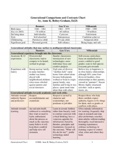
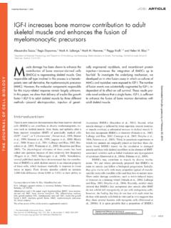
Transcription of The Muscular System Skeletal Muscle Tissue and …
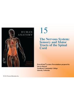
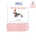
1 C h a p t e r9 The Muscular System Skeletal Muscle Tissue and OrganizationPowerPoint Lecture Slides prepared by Jason LaPresNorth Harris CollegeHouston, TexasCopyright 2009 Pearson Education, Inc.,publishing as Pearson Benjamin CummingsIntroduction Humans rely on muscles for many of our physiological processes, and virtually all our dynamic interactions with the environment involve Muscle Tissue . Copyright 2009 Pearson Education, Inc., publishing as Pearson Benjamin CummingsMuscles and LifeIntroduction There are three types of Muscle Tissue : Skeletal Muscle Skeletal Muscle Tissue moves the body by pulling on bones of the skeleton. Cardiac Muscle Cardiac Muscle Tissue pushes blood through the arteries and veins of the circulatory System .
2 Smooth Muscle Smooth Muscle tissues push fluids and solids along the digestive tract and perform varied functions in other systems. Copyright 2009 Pearson Education, Inc., publishing as Pearson Benjamin CummingsIntroduction Muscle tissues share four basic properties: Excitability: the ability to respond to stimulation Skeletal muscles normally respond to stimulation by the nervous System . Cardiac and smooth muscles respond to the nervous System and circulating hormones. Contractility: the ability to shorten actively and exert a pull or tension that can be harnessed by connective tissues Extensibility: the ability to continue to contract over a range of resting lengths Elasticity: the ability of a Muscle to rebound toward its original length after a contraction Copyright 2009 Pearson Education, Inc.
3 , publishing as Pearson Benjamin CummingsFunctions of Skeletal Muscle Skeletal muscles are contractile organs directly or indirectly attached to bones of the skeleton. Skeletal muscles perform the following functions: Produce Skeletal movement Maintain posture and body position Support soft tissues Regulate entering and exiting of material Maintain body temperatureCopyright 2009 Pearson Education, Inc., publishing as Pearson Benjamin CummingsAnatomy of Skeletal MusclesFigure Organization of Skeletal MuscleCopyright 2009 Pearson Education, Inc., publishing as Pearson Benjamin CummingsAnatomy of Skeletal MusclesFigure Muscle InnervationCopyright 2009 Pearson Education, Inc., publishing as Pearson Benjamin CummingsFigure Formation and Structure of a Skeletal Muscle FiberAnatomy of Skeletal MusclesCopyright 2009 Pearson Education, Inc.
4 , publishing as Pearson Benjamin CummingsAnatomy of Skeletal MusclesFigure StructureCopyright 2009 Pearson Education, Inc., publishing as Pearson Benjamin CummingsSarcomereCopyright 2009 Pearson Education, Inc., publishing as Pearson Benjamin CummingsAnatomy of Skeletal MusclesSarcomere Structure Sarcomere Organization Thickand thin filamentswithin a myofibrilare organized in the sarcomeres. All of the myofibrils are arranged parallel to the long axis of the cell, with their sarcomeres lying side by side. Anatomy of Skeletal MusclesFigure of Functional Organization in a Skeletal Muscle FiberCopyright 2009 Pearson Education, Inc., publishing as Pearson Benjamin CummingsLayers of a MuscleCopyright 2009 Pearson Education, Inc.
5 , publishing as Pearson Benjamin CummingsAnatomy of Skeletal MusclesAnatomy of Muscle Review Layers of a Muscle Breakdown Skeletal Muscle from large to small MCopyright 2009 Pearson Education, Inc., publishing as Pearson Benjamin CummingsAnatomy of Skeletal MusclesThick Filament Thin and Thick Filaments Each thin filament consists of a twisted strand of several interacting proteins 5 6 nm in diameter and 1 m in length. Troponinholds the tropomyosinstrand in place. Thick filaments are 10 12 nm in diameter and m in length, making them much larger than thin filaments. Thin FilamentTroponinAnatomy of Skeletal MusclesFigure and Thick FilamentsCopyright 2009 Pearson Education, Inc., publishing as Pearson Benjamin CummingsMuscle Contraction Contracting Muscle fibers exert a pull, or tension, and shorten in length.
6 Caused by interactions between thick and thin filaments in each sarcomere Triggered by presence of calcium ions Contraction itself requires the presence of 2009 Pearson Education, Inc., publishing as Pearson Benjamin CummingsMuscle ContractionMuscle Contraction The Sliding Filament Theory Explains the following changes that occur between thick and thin filaments during contraction: The H band and I band get smaller. The zone of overlap gets larger. The Z lines move closer together. The width of the A band remains constant throughout the contraction. Copyright 2009 Pearson Education, Inc., publishing as Pearson Benjamin CummingsMuscle ContractionFigure in the Appearance of a Sarcomere during Contraction of a Skeletal Muscle FiberCopyright 2009 Pearson Education, Inc.
7 , publishing as Pearson Benjamin CummingsMuscle ContractionFigure Effect of Sarcomere Length on TensionCopyright 2009 Pearson Education, Inc., publishing as Pearson Benjamin CummingsMCopyright 2009 Pearson Education, Inc., publishing as Pearson Benjamin CummingsMuscle ContractionCalcium and Troponin Interaction The Start of a Contraction Triggered by calcium ions in the sarcoplasm Electrical events at the sarcolemmal surface Trigger the release of calcium ions from the terminal cisternae The calcium ions diffuse into the zone of overlap and bind to troponin. Troponin changes shape, alters the position of the tropomyosin strand, and exposes the active sites on the actin molecules. Muscle ContractionFigure Orientation of the Sarcoplasmic Reticulum, T Tubules, and Individual SarcomeresCopyright 2009 Pearson Education, Inc.
8 , publishing as Pearson Benjamin CummingsMuscle Contraction The End of a Contraction When electrical stimulation ends: The SR will recapture the Ca2+ions. The troponin tropomyosin complex will cover the active sites. And, the contraction will 2009 Pearson Education, Inc., publishing as Pearson Benjamin CummingsMuscle ContractionFigure Neuromuscular SynapseCopyright 2009 Pearson Education, Inc., publishing as Pearson Benjamin CummingsMuscle ContractionFigure Events in Muscle ContractionCopyright 2009 Pearson Education, Inc., publishing as Pearson Benjamin CummingsMotor Units and Muscle ControlFigure Arrangement of Motor Units in a Skeletal MuscleCopyright 2009 Pearson Education, Inc.
9 , publishing as Pearson Benjamin CummingsMotor Units and Muscle Control Muscle Tone Some of the motor units of muscles are always contracting, producing a resting tension in a Skeletal Muscle that is called Muscle tone. Resting Muscle tone stabilizes the position of bones and joints. Copyright 2009 Pearson Education, Inc., publishing as Pearson Benjamin CummingsMotor Units and Muscle Control Muscle Hypertrophy and Atrophy Exercise causes increases in Number of mitochondria Concentration of glycolytic enzymes Glycogen reserves Myofibrils Each myofibril contains a larger number of thick and thin filaments. The net effect is an enlargement, or hypertrophy, of the stimulated Muscle . Disuse of a Muscle results in the opposite, called 2009 Pearson Education, Inc.
10 , publishing as Pearson Benjamin CummingsTypes of Skeletal Muscle FibersCopyright 2009 Pearson Education, Inc., publishing as Pearson Benjamin CummingsTypes of Skeletal Muscle Fibers The features of fast fibers, or white fibers, are: Large in diameter due to many densely packed myofibrils Large glycogen reserves Relatively few mitochondria Their mitochondria are unable to meet the demand. Fatigue easily Can contract in seconds or less following stimulationCopyright 2009 Pearson Education, Inc., publishing as Pearson Benjamin CummingsTypes of Skeletal Muscle Fibers Slow fibers, or red fibers, features are Only about half the diameter of fast fibers Take three times as long to contract after stimulation Contain abundant mitochondria Use aerobic metabolism Have a more extensive network of capillaries than do muscles dominated by fast Muscle fibers.