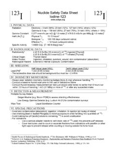
Transcription of THYROID IMAGING STUDY (I-123 as Sodium Iodide)
1 Reviewed/ 48-1 THYROID IMAGING STUDY - I-123 Revised: 6/26/2006 THYROID IMAGING STUDY (I-123 as Sodium iodide ) Overview The THYROID IMAGING STUDY with radioiodine demonstrates the distribution of functioning THYROID tissue, including ectopic tissue, since THYROID tissue is the only tissue that concentrates large amounts of iodine. Indications Evaluation of hyperthyroidism (1-3). Evaluation of enlarged glands or glands with nodules (1-3). Evaluation of patients who had irradiation of the head and neck in childhood with or without palpable nodules (4,5). Evaluation for ectopic THYROID tissue, struma ovarii (image over pelvis) and lingual THYROID (image upper neck and jaw). Evaluation of congenital hypothyroidism (6). Examination Time Initially: 20 minutes for radiopharmaceutical administration. IMAGING at 6 hours: 30 minutes. Patient Preparation (7) The patient must be off THYROID hormones: 1. Thyroxine (T-4) for at least 10 days. 2. Triiodothyronine (T-3) for at least 3 days.
2 The patient must not be taking antithyroid medications: 1. Propylthiouracil (PTU) and tapazole for at least 3 days. The patient must not have had intravenous or intrathecal iodinated contrast material (IVP, CT with contrast, myelogram, angiogram) for at least 3 weeks. The technologist records a pertinent, standard history on the THYROID Information Sheet (see below). The nuclear medicine physician records his/her palpation findings on the same form. Reviewed/ Revised: 6/26/2006 THYROID IMAGING STUDY - I-123 48-2 Equipment & Energy Windows Gamma camera: Small or large field of view. Collimator: Pinhole with 5 mm insert. Energy windows: 20% window centered at 159 keV. Radiopharmaceutical, Dose, & Technique of Administration Radiopharmaceutical: I-123 as Sodium iodide (8-10). Dose: mCi ( MBq). Technique of administration: Oral. Patient Position & IMAGING Field Patient position: Supine with the chin tilted up. IMAGING field: Neck. Acquisition Protocol Begin IMAGING 6 hours after ingestion of the radiopharmaceutical.
3 Acquire a 5 minute ANT image of the THYROID with the collimator 6 cm from the patient s neck and with a 4 cm radioactive ruler in the field of view (11): 1. A 6 cm long block can be made as a convenient measuring device. 2. A 4 cm ruler can be constructed by marking a 4 cm distance on a 5 cm long strip of aluminum. Drops of pertechnetate are then placed at the 4 cm marks and allowed to dry. 3. The strip is place vertically in the midline so that the inferior marker is over the suprasternal notch. 4. Usually the activity in the drops of pertechnetate is high enough that the markers are visible through the I-123 energy window. Acquire a second 5 minute ANT image with the distance between the collimator and patient s neck adjusted so that the THYROID gland fills three quarters of the field of view. Use the persistence scope to determine this distance. Acquire 5 minute RAO and LAO oblique images at 35 ; again with the THYROID gland filling approximately three quarters of the field of view (12).
4 Reviewed/ 48-3 THYROID IMAGING STUDY - I-123 Revised: 6/26/2006 If there is a palpable nodule, an additional 5 minute ANT image should be acquired: 1. The nuclear medicine physician places a mark on the patient s skin directly over the center of the palpable nodule. 2. The technologist then places a radioactive marker on top of the skin mark. 3. An image may now be exposed or, if the camera is equipped with an electronic marker, the electronic marker can be aligned with the radioactive marker, the radioactive marker removed, and then an image exposed. 4. In either case, the camera must be positioned so that its central ray (a line perpendicular to the crystal and passing through the pinhole) passes through the palpable nodule (13). Protocol Summary Diagram I-123 Sodium iodide Action Delayed pinhole images Time 0 6 hr hr Data Processing None.
5 Optional Maneuvers Evaluation of midline activity: If the images show midline radioactivity which may be due to radioactive saliva, have the patient swallow water and repeat the image. IMAGING of suppressed THYROID tissue with Tl-201 (14): 1. Perform at least 1 day after I-123 STUDY . 2. Inject 2 mCi thallous Tl-201 chloride intravenously. 3. At 15 minutes acquire a 10 minute ANT image of THYROID using a pinhole collimator. THYROID suppression test (15): 1. Performed to determine if a functioning nodule is autonomous. 2. Place the patient on 25 g of triiodothyronine four times a day for three days and repeat the THYROID IMAGING STUDY . Reviewed/ Revised: 6/26/2006 THYROID IMAGING STUDY - I-123 48-4 Principle Radiation Emission Data - I-123 (16) Physical half-life = hours. Radiation Mean % per disintegration Mean energy (keV) Gamma-2 ce-K, gamma-2 Dosimetry - I-123 as Sodium Iodine (17,18)
6 Organ rads/500 Ci MBq THYROID Stomach wall Ovaries Red marrow Liver Whole body Testes References 1. Beierwaltes WH: Endocrine IMAGING in the management of goiter and THYROID nodules: Part I: J Nucl Med 32:1455-1461, 1991. 2. Ross DS: Evaluation of the THYROID nodule. J Nucl Med 32:2181-2192, 1991. 3. Intenzo CM, dePapp AE, Fabbour S, et al: Scintigraphic manifestations of thyrotoxicosis.
7 RadioGraphics 23:857-869, 2003. 4. Hancock SL, Cox RS, McDougall IR: THYROID diseases after treatment of Hodgkin s disease. New Engl J Med 325:599-605, 1991. 5. Fogelfeld L, Wiviott MBT, Shore-Freedman E, et al: Recurrence of THYROID nodules after surgical removal in patients irradiated in childhood for benign conditions. N Engl J Med 320:835-840, 1989. 6. Panoutsopoulos G, Mengreli C, Ilias I, et al: Scintigraphic evaluation of primary congenital hypothyroidism: Results of the Greek screening program. Eur J Nucl Med 28:529-533, 2001. 7. Sarkar SD: In vivo THYROID studies. In Diagnostic Nuclear Medicine, A Gottschalk, PB Hoffer, EJ Potchen, eds, Williams & Wilkins, Baltimore, 1988, pp 756-768. 8. Ryo UY, Vaidya PV, Schneider AB, et al: THYROID IMAGING agents: A comparison of I-123 and Tc-99m pertechnetate. Radiology 148:819-822, 1983. 9. Beierwaltes WH: Editorial: Comparison of technetium-99m and iodine-123 nodules: Correlation with pathologic findings. J Nucl Med 31:400-402, 1990.
8 10. Palmer DW, Rao SA: A simple method to quantitate iodine-124 contamination in iodine-123 radiopharmaceuticals. J Nucl Med 26:936-940, 1985. Reviewed/ 48-5 THYROID IMAGING STUDY - I-123 Revised: 6/26/2006 11. Blue PW, Carr JR: A method for sizing the pinhole THYROID image. J Nucl Med Tech 10:207-208, 1982. 12. Karelitz JR, Richards JB: Necessity of oblique views in evaluating the functional status of a THYROID nodule. J Nucl Med 15:782-785, 1974. 13. McKitrick WL, Park HM, Kosegi JE: Parallax error in pinhole THYROID scintigraphy: A critical consideration in the evaluation of substernal goiters. J Nucl Med 26:418-420, 1985. 14. Corstens F, Huysmans D, Kloppenborg P: Thallium-201 scintigraphy of the suppressed THYROID : An alternative for iodine-123 scanning after TSH stimulation. J Nucl Med 29:1360-1363, 1988. 15. Hurley JR, Becker DV: THYROID suppression and stimulation testing: The place of scanning in the evaluation of nodular THYROID disease. Sem Nucl Med 11:149-160, 1981.
9 16. 53-I-123. In MIRD: Radionuclide data and decay schemes, DA Weber, KF Eckerman, LT Dillman, JC Ryman, eds, Society Nuclear Medicine, New York, 1989, p 217. 17. MIRD Dose Estimate Report No. 5: Summary of current radiation dose estimates to humans from I-123, I-124, I-126, I-130, I-131, I-132 as Sodium iodide . J Nucl Med 16:857-860, 1975. 18. Baker GA, Lum DJ, Smith EM, et al: Significance of radiocontaminants in 123-I for dosimetry and scintillation camera IMAGING . J Nucl Med 17:740-743, 1976. Normal Findings > Brander A, Viikinkoske T, Nickels J, et al: THYROID gland: US screening in a random adult population. Radiology 181:683-687, 1991. > Hegedus L, Perrild H, Poulsen LR, et al: The determination of THYROID volume by ultrasound and its relationship to body weight, age, and sex in normal subjects. J Clin Endocrinol Metab 56:260-263, 1983. Reviewed/ Revised: 6/26/2006 THYROID IMAGING STUDY - I-123 48-6 THYROID INFORMATION SHEET Nuclear Medicine Department Institution_____ Name_____ ID_____ Age_____ Sex_____ Referring physician_____ Date_____ TECHNOLOGIST TO COMPLETE Did the patient notice a problem with his/her THYROID or neck?
10 Yes___ No___ Has the patient had previous THYROID problems? Yes___ No___ Has the patient had any THYROID surgery? Yes___ No___ Did the patient have radiation treatments to the face/neck as a child? Yes___ No___ Has the patient taken THYROID hormones or antithyroid medicines? Yes___ No___ Has the patient had any intravenous x-ray contrast in the last month? Yes___ No___ If the patient is having an uptake STUDY , have he/she eaten since last night? Yes___ No___ Additional pertinent THYROID history: _____ _____ Technologist_____ PHYSICIAN TO COMPLETE Neck Palpation Diagram Check as Appropriate






![F]Tetrafluoroborate ([ F]TFB) and its analogs for PET ...](/cache/preview/c/9/0/5/f/3/6/b/thumb-c905f36b196cece02703fab1700529b9.jpg)