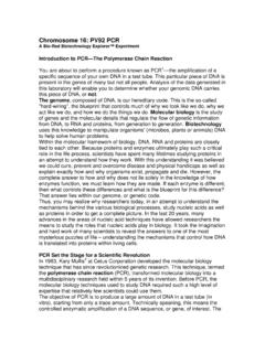
Transcription of Title: Column Chromatography of Green …
1 Minutman Regional High School Document 758 Marrett Road Revision Number 3 Lexington, MA 02420 Effective Date: 09 Jul13 title : Column Chromatography of Green fluorescent Protein Approvals: Preparer Date_07 Oct06_____Reviewer: Mary Jane Kurtz _____Date____09 Jul13_____ Page 1 of 6 Part I Crude Isolation of GFP from Lysed Cells q 1. Purpose: The purpose of this SOP is to purify Green fluorescent protein (GFP) from lysed E. coli cells using Column Chromatography . 2. Scope: This SOP will be executed whenever the purification of GFP is needed.
2 This SOP will be executed after the performance of pGLO Bacterial Transformation. 3. Responsibility: It is the responsibility of the Instructor to aid the students in the performance of all the procedures described in this SOP. It is the responsibility of the Instructor to update this SOP as needed. 4. References: Instructions from biotechnology explorer Green fluorescent Protein (GFP) Purification Kit Instruction Manual (Bio-Rad catalog number 166-0005 EDU GFP Chromatography Kit. (Bio-Rad catalog number 166-0005 EDU) 5. Definitions: Ion Exchange Chromatography Page 2 of 6 Minutman Regional High School Document 758 Marrett Road Revision Number 3 Lexington, MA 02420 Effective Date: 09 Jul13 Hydrophobic Interaction Chromatography Elution Eluate Column Supernatant 6.)
3 Hazard Communication Wear personal protection equipment Wear eye protection. For Disposal of Biohazard Waste use an autoclavable bag and follow proper sterilization techniques. 7. Materials Safety glasses or goggles Microfuge tubes Pipettes Microtube rack Marker E. coli GFP cell culture from Bacterial Transformation and Upstream Processing SOP Incubator to grow E. coli transformed with GFP at 35 C Freezer Microfuge centrifuge TE Buffer Lysozyme in 1ml of TE Buffer UV Lamp 8. Procedure: Preliminary Procedures before Chromatography Take 2ml of pGLO transformed culture from the 20ml culture tube, centrifuge at 2,000 RPM for twenty minutes. The cell pellet should fluoresce when viewed by a UV lamp.
4 While waiting, rehydrolyze the lysozyme with 1 ml of TE buffer. Pour off the E. coli supernatant into a 10% Clorox solution. Add 750 l of TE buffer and resuspend the pellet by pipetting the fluid up and down many times to make a homogenous mixture. Vortex if necessary. Add 1 drops of the reconstituted lysozyme solution. Mix the contents Page 3 of 6 gently. Freeze contents at -20 C for 1 day at least. Thaw the microfuge tube containing bacterial cell lysate to hand warmth temperature. Minutman Regional High School Document 758 Marrett Road Revision Number 3 Lexington, MA 02420 Effective Date: 09 Jul13 Centrifuge for 10 minutes at maximum speed.
5 Observe the bacterial cell supernatant with the UV light. It should fluoresce. Remove the bacterial cell lysate supernatant and place into a clean microfuge tube. This is the fraction we will use to purify GFP. Part II Hydrophobic Interaction Chromatography Materials 250ul of bacterial cell lysate Pre-packed Hydrophobic Interaction Chromatography Column Waste tube or beaker HIC Buffers: Binding buffer (= ammonium sulfate in TE Buffer pH 8) Equilibration buffer (= ammonium sulfate in TE Buffer pH8) Wash buffer (= ammonium sulfate in TE Buffer pH8) Elution buffer (= TE buffer) Test tube rack Magic marker 3 test tubes (5 ml volume) disposable pipettes ~ 3 ml Pipetman 1000ul with tips UV black light Procedure (Illustrated Overview).
6 Page 4 of 6 Remove caps from the top and bottom of the HIC Column and then allow the fluid within to drain. Minutman Regional High School Document 758 Marrett Road Revision Number 3 Lexington, MA 02420 Effective Date: 09 Jul13 Add 2 ml of Equilibration buffer to the top of the Column . Allow the Column to drain into the waste beaker. Cap the bottom of the Column . Transfer 250 l of bacterial cell lysate to a microfuge tube and add 250 l of binding buffer to it. Label three 5ml plastic test tubes in sequence from 1-3.
7 Place test tube number 1 under the Column . Remove the cap from the bottom of the Column and add 250 l of the bacterial cell lysate supernatant/ Binding buffer used in to the top of the Column . Allow the solution to drain completely into test tube 1. Place test tube number 2 under the Column . Add 250 l of Wash buffer. Allow the buffer to drain to the top of the Column into test tube 2. Place test tube number 3 under the Column . Add 750 l of Elution buffer to the Column . Allow the buffer to drain completely into test tube 3. This tube should have pure GFP in it. It can be seen glowing Green under the long range UV lamp.
8 Note the GFP positive test tube(s) and save for future analysis by SDS-PAGE. Part III Ion Exchange (Anion Ion Exchange) Chromatography (IEX) Materials: 250 l of bacterial cell lysate supernatant Pre-packed MacroPrep High Q for Anion Exchange Chromatography Column Waste tube or beaker Anion Ion Exchange Chromatography buffers: Equilibration buffer (=50 mM Tris pH ) Elution buffer 1 (50 mM Tris, pH , 130 mM NaCl) Elution buffer 2 (50 mM Tris, pH , 200 mM NaCl) Elution buffer 3 (50 mM Tris pH , 300 mM NaCl) Elution buffer 4 (50mM Tris pH , 500 mM NaCl) Test tube rack Marker 6 test tubes (5 ml volume) Disposable pipettes ~ 3 ml Pipetman 1000ul with tips UV black light Lab marker Microfuge tube 12.
9 Procedure: Label 5, 5ml test tubes in sequence from 1-5. Remove cap from the top and then the bottom of the IEX Column Page 5 of 6 Minutman Regional High School Document 758 Marrett Road Revision Number 3 Lexington, MA 02420 Effective Date: 09 Jul13 Equilibrate the Column by adding 2ml of 50mM Tris pH Collect and discard into the waste beaker. Load 250 l of the bacterial cell lysate supernatant onto the top of the Column and collect the flow through fraction in test tube 1.
10 Examine with UV light. Save 10ul of the flow through in a microfuge tube for future use. Immediately add 250ul of Elution Buffer 1 (50 mM Tris, pH , 130 mM NaCl) to the top of the Column and collect the eluate in test tube 2. Examine with UV light. Save 10ul of the flow through in a microfuge tube for future use. Add 250ul of Elution Buffer 2 (50 mM Tris, pH , 200 mM NaCl) to the top of the Column and collect the eluate in test tube 3. Examine with UV light. Save 10ul of the flow through in a microfuge tube for future use. Add 750ul of Elution Buffer 3 (50 mM Tris, pH , 300 mM NaCl) to the top of the Column and collect the eluate in test tube 4. Examine with UV light. Save 10ul of the flow through in a microfuge tube for future use.