Transcription of Wagner Cone Prosthesis® Hip System ... - Zimmer …
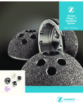
1 Wagner Cone prosthesis . Hip Stem Surgical Technique 1 08:44:08. Wagner Cone prosthesis Hip Stem Surgical Technique 3. Surgical Technique Table of Contents Wagner Cone prosthesis History 4. Design Features 4. Wagner Cone prosthesis 5. Instruments 6. Indications and Contraindications 7. Preoperative Planning 8. Surgical Technique 9. Postoperative Treatment 18. Case Studies 19. Implants 20. Instruments 21. Literature 24. 2-3 08:44:08. 4 Wagner Cone prosthesis Hip Stem Surgical Technique Wagner Cone prosthesis Hip Stem Surgical Technique 5. History Design Features Wagner Cone prosthesis Hip Stem 1990: First implantation Uncemented implant with free The Wagner Cone prosthesis Stem is In addition to providing rotational sta- As the CCD angle and the offset are 1992: Market introduction setting of antetorsion designed for uncemented fixation in bility, the sharp longitudinal ribs of not constant values, the Wagner 2006: Slight design modification Designed for difficult bone difficult bone conditions at the proxi mal the stem are also beneficial for bony Cone prosthesis Stem is now available (no design change on the zone of conditions at the proximal end of end of the femur and for CDH cases.)
2 Apposition. Schenk's1 investigations in 2 different CCD angles, 125 and primary fixation): the femur and for CDH cases have shown very clearly that bone forms 135 . This provides a wider range of Enlarged proximal shoulder with Tapered shape with an angle of The surface of the prosthesis is rough- and attaches preferentially on the offset options, which allows an ribs till the tip of the shoulder 5 degrees for press-fit fixation blasted which, together with the sharp-edged prominences of the implant adequate restoration of biomechanical for a bigger surface of osseo- 8 longitudinal ribs for rotational characteristic shape, promotes bony and less in the hollows of the surface. parameters, as the center of rota- integration stability apposition over a large tion, the CCD angle and the leg length. Slim neck and short cone for a Standard and offset version for a In order to achieve a broad-based better range of motion better restoration of the anatomy The circular tapered stem is not subject support of the prosthesis in the region Both versions are available in 12 diam- 2006: Additional offset version 125 to any rotation force during inser- of the calcar, the medial rib is insert- eters from 13 to 24 mm to fit the to better restore the anatomy tion, i.
3 E. the angle of antetorsion can ed distally to this area into the convex individual width of the medullary canal. be determined by the surgeon. The support surface. The ribs on the later- stem has 8 longitudinal ribs. The rela- al part start from the tip of the shoulder tively sharp ridges of the ribs cut in order to ensure the greatest pos- into the bone, thus allowing for optimal sible area of contact in the trochanter. rotational This also explains This provides rotational stability and the fact that the typical thigh pain improves osseointegration. associated with some uncemented prosthetic systems is practically unknown with the Cone Prosthesis3. 1990 2006 From 2006 Wagner Cone prosthesis 125 and 135 The round stem facilitates unimpeded rotation during implantation to adjust the antetorsion angle. 1. Schenk , Wehrli U. Zur Reaktion des Knochens auf eine zementfreie SL-Femur-Revisionsprothese. Orthopade. 1989; 18: 454 462. 2. B hler D., Berlemann U, Lippuner K, Jaeger P, Nolte L.
4 Three-dimensional primary stability of cementless femoral stems. Clinical Biomechanics. 1997; 12: 75 85. 3. Wagner H., Wagner M. Cone prosthesis for the hip joint. Arch Orthop Trauma Surg. 2000; 120: 88 95. 4-5 08:44:18. 6 Wagner Cone prosthesis Hip Stem Surgical Technique Wagner Cone prosthesis Hip Stem Surgical Technique 7. Instruments Indications and Contraindications Surgeons have a set of user-friendly Modular Trial Stems Indications 2. Increased antetorsion of the Contraindications instruments at their disposal Trial stems are used to determine the The Wagner Cone prosthesis Hip Stem femoral neck, e. g. CDH, where the The first contraindication is the for implantation of the Wagner Cone exact positioning and correct size of the is designed for cylindrical proximal cross-section of the proximal trumpet-shaped proximal widening of prosthesis Stem. The core instruments final implant. The trial stems of the femoral bone that could fracture when medullary cavity is anteverted even the medullary cavity where no support are reamers which are used for the Wagner Cone prosthesis Implant are conventional flared stems are used.
5 After resection of the femoral for the prosthetic stem in its middle and careful preparation of the medul- modular with one distal part for each The Wagner Cone prosthesis Stem is neck, forcing prosthetic stems with proximal thirds is available. In addition lary canal as well as modular trial size and two different proximal parts, also designed for deformities of the a flat or transverse oval profile the Wagner Cone prosthesis Stem stems used for determining the 125 and 135 , per size. This allows femur where fixation of conventional into antetorsion. With the circular should not be considered when there most adapted Wagner Cone prosthesis maximal intra-operative flexibility stems is problematic. Specifically, cross section of the Wagner is considerable weakening of the bone Stem version and the best-suited size during trial reduction. The proximal part the Wagner Cone prosthesis Stem is Cone prosthesis Stem, the angle structure at the proximal end of the of implant.
6 Can be exchanged in situ leaving the indicated when the following factors of antetorsion can be adjusted femur. distal part in the bone to avoid damage are present: as desired. to the bone. Reamers 1. Cylindrical configuration of the proxi- 3. Deformities and intramedullary The Wagner Cone prosthesis Stem, with Please note that the distal parts of the mal medullary cavity where inser- bony scarring of the proximal end its circular cross-section, is indicated trial stems have only four ribs (instead tion of conventionally shaped of the femur after osteotomies, particularly for slender configurations of eight, as on the implants) to facil- prosthetic stems causes problems. fractures and growth disorders or of the proximal femur, as well as for itate the extraction of the trial stem These conditions are found espe- congenital deformities. Preparing cases involving pathologic morphology. and to avoid unnecessary damage cially with congenital dysplasia of the medullary cavity to receive When these indications are present, to the bone during the trial procedure.
7 The hip (CDH) and coxa vara con- the prosthesis with conventional an important focus should be placed The trial stem may be inserted as far genita; they are also found with a reamers can be very tedious or on the preparation of the medul- as the intended anchoring depth of the very delicate bone structure, even impossible, whereas prepa- lary canal in the most bone-preserving implant to assess accurately the ten- frequently in combination with coxa ration with the conical reamer of manner. The use of reamers allows sion of the soft tissue, range of motion valga. the Wagner Cone prosthesis Stem the surgeon to prepare the medullary and degree of antetorsion. can be carried out more easily and canal with care and precision, even safely. in cases of poor bone quality of the proximal femur where the use of rasps is not an option. Reamer Trial Stem 6-7 08:44:26. 8 Wagner Cone prosthesis Hip Stem Surgical Technique Wagner Cone prosthesis Hip Stem Surgical Technique 9. Preoperative Planning Surgical Technique The preoperative planning follows In choosing the diameter, it must be Using the X-ray template, the outline of The Wagner Cone prosthesis Hip Stem The incision or resection of the posterior standard procedures.
8 A good-quality remembered that reaming with the the selected Wagner Cone prosthesis can be implanted using all of the usual joint capsule is a critical issue in the X-ray template is essential. The first reamer removes a thin layer of bone Stem is then transferred to the planning surgical approaches. However, posterior posterior approach. Posterior dislocation step in planning consists in selecting and that the sharp longitudinal ribs sketch and the line of resection is also access with the patient in the lateral of the prosthesis can occur more suitable implants for the acetabulum cut slightly into the bone during inser- drawn from the template. The plan- position is particularly suitable: the readily during the healing phase, if the and the femur using the X-ray templates tion. The outline of the prosthetic ning sketch is now laid on the X-ray film reamer for the Wagner Cone prosthesis cup and/or the stem are placed in on the original X-ray films. stem on the planning template must and the outline of the femur is trans- Implant has a straight stem which is insufficient anteversion.
9 Therefore overlap the inner outline ferred carefully. The tip of the greater introduced into the axis of the medullary When selecting the size of the Wagner of the cortex in the region of the middle trochanter on the X-ray is at the level cavity. With the posterior approach, This problem can be minimized with Cone prosthesis Stem it is important third of the stem by 1 mm on each side. of the previously made marking for the when the hip and knee are flexed, the a trial reduction with the trial stems that the configuration of the femur desired trochanter position. way to the medullary cavity is free before definitive implantation of the allows close contact between the Furthermore, the most adapted version without the need for temporary removal Wagner Cone prosthesis Hip Implant. middle third of the prosthetic stem of the Wagner Cone prosthesis Stem Finally, the distance between the of the greater trochanter and without But nonetheless, this phenomen and the cortex, and not just that (125 or 135 ) to best restore the offset, cone of the stem and the proximal limit the instrument exerting pressure on the requires particular care.
10 The tip of the stem fits tightly in the the center of rotation and the CCD angle of the lesser trochanter is measured. muscles. medullary cavity. is selected. In reconstructing deformed hip joints, further marking points can be given With the lateral, transgluteal and Selection of the correct stem diameter Planning drawing: On the outline draw- and their distances measured. All longi- anterior approaches, retraction is particularly important. The most ing of the pelvis, the position of the tudinal measurements must be made of the muscles is more difficult. More- common mistake consists in choosing cup implant with the center of rotation according to the scale on the template over, with the posterior approach, a stem diameter that is too thin. Such is first sketched and the current and as this takes into account the degree the incision is smaller and there is less a decision can result in secondary sub- desired position of the tip of the tro- of magnification of the X-ray.