Transcription of Laboratory 8 - Urinary and Reproductive Systems
1 Laboratory 8 - Urinary and Reproductive SystemsUrinary SystemPlease read before starting: It is easy to damage the structures of the Reproductive systemas you expose structures associated with excretion, so exercise caution as you do also note that we will have drawings available as well to help you find and identifythe structures described major blood vesselsserving the kidneys are therenal artery and the renalvein., which are located deepin the parietal renal artery is a branch ofthe dorsal aorta that comes offfurther caudal than the cranialmesenteric the left kidney in situ,dividing it into dorsal andventral portions by making afrontal section along the outerperiphery.
2 Observe the renal cortexrenal medulla (next layer in)renal pyramidsrenal pelvisureter (see above diagram)The kidneys include a variety of structures including anarterial supply, a venous return, extensive capillarynetworks around each nephron and then, of course, thefiltration and reabsorption apparatus. These structures areprimarily composed of nephrons (the basic functional unitof the kidney) and the ducts which carry urine away fromthe nephron (the collecting ducts and larger ductseventually draining these into the ureters from eachkidney. The renal pyramids contain the extensions of thenephrons into the renal medulla (the Loops of Henle) and the collecting ducts.)
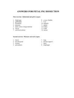
3 Urine iseventually emptied into the renal pelvis before leaving the kidneys in the ureters leaves the kidneys medially at approximately the midpoint of the organs andthen run caudal to the Urinary bladder. The Urinary bladder is a longish bag that liesbetween the umbilical you pull on the pigs umbilicalcord, you will extend the urinarybladder and make it easier tolocate the urethra. The urethraleads into the penis in males andinto the vagina in ReproductiveAnatomy: If your pig isfemale, observe these structuresin a male pig being dissected byanother two figures to the rightshow the male reproductivesystem dissected so that themajor structures are penis is located in the flapof ventral body wall caudal tothe umbilical cord.
4 Make alengthwise incision using yourscissors and beginning at theurogenital opening to expose thepenis. Make sure to cut onlyjust below the skin. Once youhave done this, you can use theprobe from your kit to exposethe penis. It will extend towardsthe back of the animal untilmeeting the in the adult male will becarried in the ductus deferentia(singular: ductus deferens),which extend from theepididymis and testes then pass over the ureters before entering the urethra. The ductusdeferentia extend backwards and toward the exterior of the animal where they passthrough the inguinal canal and into the scrotum.
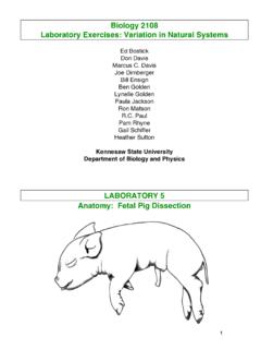
5 The testis can be exposed by cuttingalong the inguinal canal (scissors are best for this). Inside the membranes housing thetestis, you should also find the connection between the ductus deferentia and theepididymis .Locate the pubic symphysis, a portion of the pelvic girdle, by probing through the muscleand connective tissue in approximately the location shown in the upper picture abovewith the dotted line. Using scissors, cut through the bone once you locate it, going fromposterior to anterior and being careful to cut only the bone. Press the hind limbs apartand trim the ends of the symphysis with your scissors.
6 This should make the urethravisible. Follow the urethra to the Urinary bladder and you should also see two largebulbourethral glands to either side of the urethra near where it meets the penis. Theseglands are also known as 'Cowper's glands' and help to protect sperm in the acidicenvironment of the urethra by producing an alkaline on the umbilical cord helps to locate the seminal vesicles. These glands arefound on the dorsal surface of the urethra very near where the ductus deferentia join theurethra. The prostate gland lies between the lobes of the seminal vesicles but will bedifficult to identify at this immature stage of development.
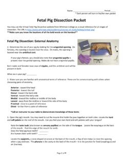
7 In adulthood, this glandproduces fluid in which the sperm are : What is the functional reason the testis very often found in an external scrotumin mammals?Female Reproductive Anatomy:As with the male discussedabove, locate the pubicsynthesis and trim as with themale. The ovaries are foundin the abdominal cavity theends of the uterine uterine tubes also go bythe more familiar names ofoviducts and fallopian tubesand are the usual site offertilization (see figure toright here).Moving down from the ovary, the uterine tubes connect to the horns of the uterus whichconnect to the body of the uterus (found above the urethra - see figure below).
8 Continuing to move caudal,the next portion of the uterusis the cervix. The cervix leadsinto the vagina. To see thecervix and vagina, dissect theuterus open with a scalpel,then look for a series ofinternal ridges (present in thecervix, but lacking in thevagina). The next structureleading externally is thevaginal vestibule, foundwhere the vagina and urethrameet. The vaginal vestibuleleads to the exterior orifice,urogenital opening, foundbelow the anus on the exteriorof the Pig UterusWe will have a pregnant pig uterus on demonstration and your will dissect anembryo from the extraembryonic membranes that support its development.
9 Theseinclude the serosa, the allantois and the amnion. The amnion surrounds the developingfetal pig. The allantois is an outpouching of the gut that plays the major role in exchangebetween the mother and fetus. The serosa is the membrane facing into the lumen of theuterus. Together, the serosa and the allantois form the chorion. The chorion forms villiwhich penetrate into the lining of the uterus (this lining is maternally derived) forming aclose relationship and highly vascularized exchange structure. The combined chorionand uterine mucosa in the area of contact are referred to as the placenta.