Thin Blood Smears
Found 11 free book(s)
Diagnosis and Treatment of Malaria in India
nvbdcp.gov.inMicroscopy of stained thick and thin blood smears remains the gold standard for confirmation of diagnosis of malaria. The advantages of microscopy are: • The sensitivity is high. It is possible to detect malarial parasites at low densities. It also helps to quantify the parasite load. • It is possible to distinguish the various species of ...
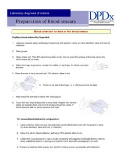
Blood collection for thick or thin blood smears
www.cdc.govMaking thick and thin blood smears . 1. Whenever possible, use separate slides for thick and thin smears. 2. Thin film (a): Bring a clean spreader slide, held at a 45° angle, toward the drop of blood on the specimen slide. 3. Thin film (b): Wait until the blood spreads along the entire width of the spreader slide. 4.
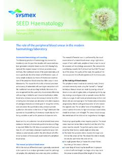
SEED Haematology – The role of the peripheral blood smear ...
www.sysmex-europe.comBlood smears that are too thin or too thick present a prob-lem. Extremely thin smears (caused by too small a drop, too slow spreading or too low a spreader angle), may result in red blood cells (RBC) that appear as spherocytes and in- creased white blood cells (WBC), such as monocytes and neutrophils, in the tails.
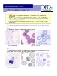
Plasmodium malariae - Centers for Disease Control and ...
www.cdc.govB. Blood smears, at least two thick and two thin, should be prepared as soon as possible after collection. Delay in preparation of the smears can result in changes in parasite morphol-ogy and staining characteristics. C. Schüffner’s dots can be demonstrated in Giemsa stain, which is preferred to Wright or Wright-

LABORATORY MANUAL FOR LABORATORY TECHNICIAN …
www.shoklo-unit.comspecimen especially thin and thick blood smear in case the lab technicians cannot do it by their own. Bad smears can lead to wrong results, so lab technician supervisor should show the medics and nurses how to make a good smear and explain that the quality of the result depends on it. However, blood
BLOOD SMEAR BASICS - NC State Veterinary Medicine
cvm.ncsu.eduthin smears that lack a dense body, thin monolayer and well-developed feathered edge. The leading edge may have a line of blood instead of a thin, feathered appearance from pushing the blood instead of pulling it. Often there is streaking in the smear. Linear lines arranged horizontally to the leading edge

Blood Cell Identification Graded
webapps.cap.orgBlood Cell Identification ... common feature found in blood smears from patients with myeloproliferative neoplasms. 5 ... the nucleus exhibits segmentation with two or more lobes connected by a thin filament. An increased number of eosinophils may occur associated with drug reactions, parasitic infections, allergic conditions, ...

Practical hematology - Islamic University of Gaza
site.iugaza.edu.ps2. As soon as the drop of blood is placed on the glass slide, the smear should be made without delay. Any delay results in an abnormal distribution of the white blood cells, with many of the large white cells accumulating at the thin edge of the smear.Rouleaux of the red blood cells and platelet clumping may also occur.

Surgical Pathology
a1.mayomedicallaboratories.comSurgical Pathology Any UNLISTED specimen should be assigned to the CPT code which most closely reflects the work involved when compared to other specimens assigned to that code. The unit of service for CPT codes 88300 - 88309 is the SPECIMEN.A specimen …

Surgical Pathology
a2.mayomedicallaboratories.comSurgical Pathology Any UNLISTED specimen should be assigned to the CPT code which most closely reflects the work involved when compared to other specimens assigned to that code. The unit of service for CPT codes 88300 - 88309 is the SPECIMEN.A specimen is defined as tissue(s) that is/are submitted for

STAINS & STAINING
ndvsu.orgSTAINS AND DYES A dye is a general-purpose coloring agent, whereas a stain is used for coloring biological material. A stain is an organic compound containing a benzene ring plus a chromophore and an auxochrome group. chromophore is …