Transcription of 2.2.32. LOSS ON DRYING - uspbpep.com
1 EUROPEAN PHARMACOPOEIA Loss on dryingusing the conditions recommended by the manufacturerof the equipment. Manufacturers of SDS-PAGE equipmentmay provide gels of different surface area and running time and current/voltage may needto vary as described by the manufacturer of the apparatusin order to achieve optimum separation. Check that the bottom of the gel, stop the the gel assembly from the apparatus and separatethe glass plates. Remove the spacers, cut off and discard thestacking gel and immediately proceed with OF PROTEINS IN GELSC oomassie staining is the most common protein stainingmethod with a detection level of the order of 1 g to 10 gof protein per band. Silver staining is the most sensitivemethod for staining proteins in gels and a band containing10 ng to 100 ng can be of the steps in gel staining are done at room temperaturewith gentle shaking ( on an orbital shaker platform)in any convenient container.
2 Gloves must be worn whenstaining gels, since fingerprints will staining solution Rand allow to stand for atleast 1 h. Remove the staining the gel with a large excess ofdestaining solution the destaining solution several times, until thestained protein bands are clearly distinguishable on a clearbackground. The more thoroughly the gel is destained, thesmaller is the amount of protein that can be detected bythe method. Destaining can be speeded up by including afew grams of anion-exchange resin or a small sponge in thedestaining solution : the acid-alcohol solutions used in this proceduredo not completely fix proteins in the gel. This can leadto losses of some low-molecular-mass proteins during thestaining and destaining of thin gels. Permanent fixationis obtainable by allowing the gel to stand in a mixture of1 volume of trichloroacetic acid R, 4 volumes of methanol Rand 5 volumes of water R for 1 h before it is immersed inthe Coomassie staining solution Rand allow to stand for 1 h.
3 Remove the fixingsolution, add fresh fixing solution and incubate either forat least 1 h or overnight, if convenient. Discard the fixingsolution and wash the gel in a large excess ofwater Rfor1 h. Soak the gel for 15 min in a 1 per centV/Vsolutionofglutaraldehyde R. Wash the gel twice for 15 min in alarge excess ofwater nitratereagent Rfor 15 min, in darkness. Wash the gel three timesfor5mininalargeexcessofwater R. Immerse the gelfor about 1 min indeveloper solution Runtil satisfactorystaining has been obtained. Stop the development byincubation in theblocking solution Rfor 15 min. Rinse thegel withwater OF STAINED SDS POLYACRYLAMIDE GELSD epending on the staining method used, gels are treated ina slightly different way. For Coomassie staining, after thedestaining step, allow the gel to stand in a 100 g/l solution ofglycerol Rfor at least 2 h (overnight incubation is possible).
4 For silver staining, add to the final rinsing a step of 5 min ina 20 g/l solution ofglycerol two sheets of porous cellulose film inwater Randincubate for 5 min to 10 min. Place one of the sheets ona DRYING frame. Carefully lift the gel and place it on thecellulose film. Remove any trapped air bubbles and pour afew millilitres ofwater Raround the edges of the gel. Placethe second sheet on top and remove any trapped air the assembly of the DRYING frame. Place in an ovenor leave at room temperature until DETERMINATIONM olecular masses of proteins are determined by comparisonof their mobilities with those of several marker proteins ofknown molecular weight. Mixtures of proteins with preciselyknown molecular masses blended for uniform staining areavailable for calibrating gels.
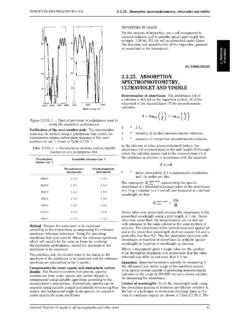
5 They are obtainable in variousmolecular mass ranges. Concentrated stock solutionsof proteins of known molecular mass are diluted in theappropriate sample buffer and loaded on the same gel as theprotein sample to be after the gel has been run, the position ofthe bromophenol blue tracking dye is marked to identifythe leading edge of the electrophoretic ion front. This canbe done by cutting notches in the edges of the gel or byinserting a needle soaked in India ink into the gel at thedye front. After staining, measure the migration distancesof each protein band (markers and unknowns) from thetop of the resolving gel. Divide the migration distance ofeach protein by the distance travelled by the tracking normalised migration distances so obtained are calledthe relative mobilities of the proteins (relative to the dyefront) and conventionally denoted the logarithm of the relative molecular masses (Mr)ofthe protein standards as a function of theRFvalues.
6 Notethat the graphs are slightly sigmoid. Unknown molecularmasses can be estimated by linear regression analysis orinterpolation from the curves of logMragainstRFas long asthe values obtained for the unknown samples are positionedalong the linear part of the OF THE TESTThe test is not valid unless the proteins of the molecularmass marker are distributed along 80 per cent of the lengthof the gel and over the required separation range ( therange covering the product and its dimer or the productand its related impurities) the separation obtained for therelevant protein bands shows a linear relationship betweenthe logarithm of the molecular mass and theRF. Additionalvalidation requirements with respect to the solution undertest may be specified in individual OF IMPURITIESW here the impurity limit is specified in the individualmonograph, a reference solution corresponding to thatlevel of impurity should be prepared by diluting the testsolution.
7 For example, where the limit is 5 per cent, areference solution would be a 1:20 dilution of the testsolution. No impurity (any band other than the main band)in the electropherogram obtained with the test solutionmaybemoreintensethanthemainbando btainedwiththereference validated conditions impurities may be quantifiedby normalisation to the main band using an integratingdensitometer. In this case, the responses must be validatedfor LOSS ON Place the prescribed quantity of the substance tobe examined in a weighing bottle previously dried underthe conditions prescribed for the substance to be the substance to constant mass or for the prescribedtime by one of the following procedures. Where the dryingGeneralNotices(1) Nuclear magnetic resonance spectrometryEUROPEAN PHARMACOPOEIA is indicated by a single value rather than arange, DRYING is carried out at the prescribed temperature 2 ) in a desiccator : the DRYING is carried out overdiphosphorus pentoxide Rat atmospheric pressure andat room temperature;b) in vacuo : the DRYING is carried out overdiphosphoruspentoxide R, ;c) in vacuowithin a specified temperature range : thedrying is carried out overdiphosphorus pentoxide R, prescribed in the monograph;d) in an oven within a specified temperature range : thedrying is carried out in an oven within the temperaturerange prescribed in the monograph.
8 E) under high vacuum : the DRYING is carried out overdiphosphorus pentoxide Rat a pressure not kPa, at the temperature prescribed in the other conditions are prescribed, the procedure to be usedis described in full in the NUCLEAR MAGNETICRESONANCE SPECTROMETRYN uclear magnetic resonance (NMR) spectrometry is basedon the fact that nuclei such as1H,13C,19F,31 Ppossessapermanent nuclear magnetic moment. When placed inan external magnetic field (main field), they take certainwell-defined orientations with respect to the direction of thisfield which correspond to distinct energy levels. For a givenfield value, transitions between neighbouring energy levelstake place due to absorption of electromagnetic radiation ofcharacteristic wavelengths at radio determination of these frequencies may be madeeither by sequential search of the resonance conditions(continuous-wave spectrometry) or by simultaneousexcitation of all transitions with a multifrequency pulsefollowed by computer analysis of the free-induction decay ofthe irradiation emitted as the system returns to the initialstate (pulsed spectrometry).
9 Aprotonmagnetic resonance spectrum appears as a set ofsignals which correspond to protons and are characteristicof their nuclear and electronic environment within themolecule. The separation between a given signal and that ofa reference compound is called a chemical shift ( )andisexpressed in parts per million (ppm); it characterises the kindof proton in terms of electronic environment. Signals arefrequently split into groups of related peaks, called doublets,triplets, multiplets; this splitting is due to the presence ofpermanent magnetic fields emanating from adjacent nuclei,particularly from other protons within two to five valencebonds. The intensity of each signal, determined from thearea under the signal, is proportional to the number ofequivalent A nuclear magnetic resonance spectrometerfor continuous-wave spectrometry consists of a magnet,a low-frequency sweep generator, a sample holder, aradio-frequency transmitter and receiver, a recorder and anelectronic integrator.
10 A pulsed spectrometer is additionallyequipped with a pulse transmitter and a computer for theacquisition, storage and mathematical transformation of thedata into a conventional less than 60 MHz for1H. Unless otherwise prescribed,follow the instructions of the recording the spectrum, verify that:1) The resolution is equal to Hz or less by measuring thepeak width at half-height usinganadequatescaleexpansionof: either the band at ppm or at ppm of thesymmetrical multiplet of a 20 per centV/Vsolution ofdichlorobenzene Rindeuterated acetone R, orthebandat ppm of a 5 per centV/Vsolution oftetramethylsilane Rindeuterated chloroform ) The signal-to-noise ratio (S/N), measured over the rangefrom 2 ppm to 5ppmonthespectrumobtainedwitha1percentV/ Vsolution ofethylbenzene Rindeuteratedchloroform R,isatleast25 of five successive determinations from the expression.