Transcription of A. Enrichment in Alkaline Peptone Water
1 Laboratory Methods for the Diagnosis of Vibrio cholerae Centers for Disease Control and Prevention Laboratory Methods for the Diagnosis of Vibrio cholerae Centers for Disease Control and Prevention IV. ISOLATION OF VIBRIO CHOLERAE FROM FECAL SPECIMENS Although V. cholerae O1 will grow on a variety of commonly used agar media, isolation from fecal specimens is more easily accomplished with specialized media. Alkaline Peptone Water (APW) is recommended as an Enrichment broth, and thiosulfate citrate bile salts sucrose (TCBS) agar is the selective agar medium of choice for isolating V. cholerae O1. In certain instances (for example, when the patient is in very early stages of illness and is passing liquid stool), it may not be necessary to enrich specimens or use selective plating media. However, Enrichment broth and a selective plating medium should always be used with convalescent patients, suspected asymptomatic infections, environmental specimens, and whenever high numbers of competing organisms are likely to be present in the specimen.
2 A. Enrichment in Alkaline Peptone Water Vibrio spp. grow very rapidly in APW, and at 6 to 8 hours will be present in greater numbers than non-Vibrio organisms. Enrichment in APW enhances the isolation of V. cholerae O1 when few organisms are present, as in specimens from convalescent patients and asymptomatic carriers. A number of other broth media have been described for Enrichment of V. cholerae. These include Monsur s Enrichment medium which contains Trypticase, potassium tellurite, and sodium taurocholate (bile salts). A modification of APW, in which potassium tellurite is added in concentrations of 1:100,000 to 1:200,000, is sometimes used. An Enrichment medium containing a selective agent may not offer any advantage over APW if it is used with a short incubation time (6 to 8 hours). B. Selective Plating Media 1. Thiosulfate citrate bile salts sucrose agar TCBS is the medium of choice for the isolation of V.
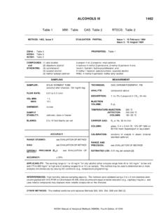
3 Cholerae and is widely used worldwide. TCBS agar is commercially available and easy to prepare, requires no autoclaving, and is highly differential and selective (Table IV-1). However, it has a relatively short shelf life once prepared (3 to 5 days) unless plates are carefully protected against drying. TCBS is subject to lot-to-lot and brand-to-brand variations in selectivity, and growth on this medium is not suitable for direct testing with V. cholerae O1 antisera. TCBS agar is green when prepared. Overnight growth (18 to 24 hours) of V. cholerae will produce large (2 to 4 mm in diameter), slightly flattened, yellow colonies with opaque centers Isolation of Vibrio cholerae from Fecal Specimens 17 | Page Laboratory Methods for the Diagnosis of Vibrio cholerae Centers for Disease Control and Prevention Figure IV-1. Overnight colonies of V. cholerae on TCBS agar are large (2-4 mm) and yellow because of the fermentation of sucrose.
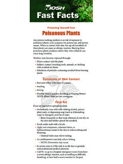
4 They are characteristically round, smooth, glistening, and slightly flattened. Figure IV-2. On TTGA medium, colonies of V. cholerae are grey, flattened, and are surrounded by a cloudy halo formed by the production of gelatinase. Isolation of Vibrio cholerae from Fecal Specimens 18 | Page Laboratory Methods for the Diagnosis of Vibrio cholerae Centers for Disease Control and Prevention and translucent peripheries (Figure IV-1). The yellow color is caused by the fermentation of sucrose in the medium. Sucrose nonfermenting organisms, such as V. parahaemolyticus, produce green to blue-green colonies. Suspicious colonies for further testing should be subcultured to a noninhibitory medium, such as gelatin agar , heart infusion agar (HIA), Kligler s iron agar (KIA), or triple sugar iron agar (TSI). 2. Taurocholate tellurite gelatin agar (TTGA or Monsur s agar ) TTGA is a selective and differential agar specifically designed for the isolation of V.
5 Cholerae. TTGA has a relatively long shelf life after preparation, and growth directly from the medium may be used for oxidase and agglutination tests (Table IV-1). The disadvantages of this medium are that it is not commercially available, and overnight colonies of V. cholerae on TTGA tend to be smaller (1 to 2 mm) than those from the TCBS agar . Potassium tellurite, which is added to the medium to increase selectivity, also varies in its quality, and each lot should be titrated to determine the optimal concentration to use in TTGA medium (see Chapter XI, Preparation of Media and Reagents). Overnight growth of V. cholerae on TTGA agar appears as small opaque colonies with slightly dark centers (Figure IV-2). After 24 hours, the centers of the colonies become darker, and eventually the entire colony becomes gunmetal grey in color. In addition to the dark coloration, which is due to the reduction of tellurite, there is also an opaque zone around colonies which resembles a halo.
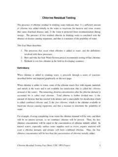
6 The halo effect, which is due to the production of the enzyme gelatinase, can be intensified by brief (15- to 30-minute) refrigeration of the plate. Because many members of the genus Vibrio have similar characteristics on TTGA, additional tests (antisera and/or biochemicals) are necessary to screen isolates from this medium. Table IV-1. Selective plating media for V. cholerae Medium Colony morphology Colony size Commercially available Autoclaved Direct testing of growth off of platea TCBS Yellow, shiny 2- 3 mm Yes No No TTGA Grey, flattened opaque zone around colony 1- 2 mm No Yes Yes MacConkeyb Colorless to light pink 1- 3 mm Yes Yes No Note: TCBS = thiosulfate citrate bile salts sucrose agar ; TTGA = taurocholate tellurite gelatin agar . a Direct testing for agglutination in antisera or oxidase reaction. b Not all strains of V. cholerae O1 will grow on MacConkey agar . Isolation of Vibrio cholerae from Fecal Specimens 19 | Page Laboratory Methods for the Diagnosis of Vibrio cholerae Centers for Disease Control and Prevention Figure IV-3.
7 Overnight growth of V. cholerae on MacConkey agar appears as small (1- to 3 mm), translucent, colorless-to-light pink (lactose-negative) colonies. 3. MacConkey (MAC agar ) MAC is used widely to isolate members of the Enterobacteriaceae and will also support the growth of some but not all strains of V. cholerae. Overnight colonies of V. cholerae on MAC tend to be small to moderately sized (1 to 3 mm) and usually appear as lactose-negative or slightly pink, often resembling colonies of late or slow lactose-fermenting organisms (Table IV-1;Figure IV-3). Suspicious colonies should be subcultured to noninhibitory media for further testing. C. Nonselective Plating Media 1. Gelatin agar (GA) GA is a good nonselective growth medium for V. cholerae. Gelatinase production, a characteristic of vibrios in general, may be determined on GA and is indicated by the production of an opaque zone around colonies which resembles a halo.
8 The halo effect can be intensified by brief (15- to 30- minute) refrigeration of the plate. Colonies of V. cholerae on GA are smooth, opaque, white, and 2-4 mm in diameter after overnight incubation at 35 to 37 C. When viewed with obliquely transmitted light with 10X to 20X magnification, colonies may appear finely Isolation of Vibrio cholerae from Fecal Specimens 20 | Page Laboratory Methods for the Diagnosis of Vibrio cholerae Centers for Disease Control and Prevention granular and iridescent with a greenish-bronze sheen. Colonies from this medium may be tested directly for agglutination with antisera as well as oxidase and string test reagents. Salt-free GA may be used as a screening medium to rule out halophilic (salt-requiring) marine vibrios resembling V. cholerae, which are frequently isolated from seafood and environmental specimens. See Chapter XI, Preparation of Media and Reagents, for special instructions for preparation of gelatin agar .
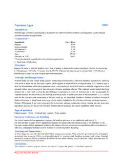
9 2. Meat extract agar (MEA, or Alkaline nutrient agar ) MEA is similar to GA in its ability to support the growth of V. cholerae. However, unlike GA, MEA does not produce colonies with differential characteristics. Colonies of V. cholerae on NEA are 2 to 4 mm in diameter after overnight incubation and are smooth, opaque, and cream colored. When viewed with oblique light with 10X to 20X magnification, colonies may appear finely granular and iridescent with a greenish bronze sheen. The oxidase test, string test and screening with antisera may be performed directly with suspicious growth on MEA plates. D. Isolation and Presumptive Identification 1. Direct inoculation of selective plating media from fecal specimens Inoculate highly selective media (TCBS, TTGA) with a heavy inoculum from liquid stool, fecal suspension, or a rectal swab (Figure IV-4). With media of low selectivity (GA, MEA, blood agar ), use a light inoculum.
10 The inoculum should be streaked with a wire loop to give a large number of isolated colonies. It is not necessary to flame the loop between streaking different quadrants on the plate. Media of high selectivity require more cross streaking into previous quadrants than media with lower selectivity. After inoculation, incubate plates for 18 to 24 hours at 35 to 37 C. 2. Inoculation of APW from fecal specimens APW can be inoculated with liquid stool, fecal suspension, or a rectal swab (Figure IV-4). The stool inoculums should not exceed 10% of the volume of the broth. Incubate the tube with the cap loosened at 35 to 37 C for 6 to 8 hours. After 6 to 8 hours incubation, subcultures to TCS should be made with one to two loopfuls of APW from the surface and topmost portion of the broth, since vibrios preferentially grow in this area. Do not shake or mix the tube before subculturing.