Transcription of AMEBIASIS - ldh.la.gov
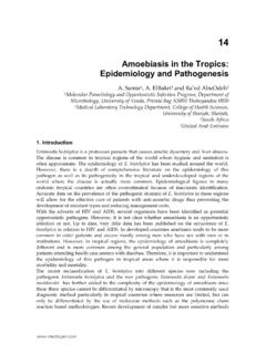
1 Louisiana Office of Public Health Infectious Disease Epidemiology Section Page 1 of 3 AMEBIASIS Revised 11/30/2011 Epidemiology AMEBIASIS is an infection caused by a protozoan parasite formerly called Entamoeba histolytica. Entamoeba histolytica has been reclassified into two species that morphologically are identical but genetically distinct. Entamoeba histolytica and Entamoeba dispar organisms are excreted as cysts or trophozoites in stools of infected persons.
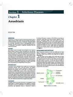
2 Infection occurs following ingestion of the cysts. Trophozoites are not infectious because they are destroyed by the acidity of the stomach and intestinal enzymes. The usual mode of transmission in the United States is by person-to-person spread and occasionally through contaminated food, drink or enema equipment. The incubation period ranges from a few days to months or years, but commonly is one to four weeks. Infectivity period: Infected patients excrete cysts intermittently, sometimes for years if untreated. Entamoeba histolytica can be found worldwide but is more prevalent in persons of lower socioeconomic status who live in developing countries where the prevalence of amebic infection may be as high as 50%.
3 The high risk groups in the US are immigrants from endemic areas, long-term visitors to endemic areas, institutionalized persons, and men who have sex with men. Clinical Description Entamoeba histolytica causes invasive disease, while E dispar is a noninvasive parasite that does not cause disease. Several clinical presentations have been associated with E. histolytica: Asymptomatic infection most likely due to E. dispar Intestinal AMEBIASIS or amebic colitis: increasing diarrhea progressing to grossly bloody dysenteric stools with lower abdominal pain and tenesmus, weight loss, fever.
4 Progressive involvement of the colon may lead to more severe complications Acute fulminant or necrotizing colitis, Ameboma: annular lesion of the cecum or ascending colon that may be mistaken for colonic carcinoma or as a tender extrahepatic mass mimicking a pyogenic abscess Liver abscess; disease is more severe in the very young, the elderly, and pregnant women. Immunosuppression, malnutrition, young age and pregnancy contribute to more severe disease. Surveillance AMEBIASIS is a not a reportable condition in Louisiana unless occurring as a suspected cluster of cases.
5 Infectious Disease Epidemiology Section Office of Public Health, Louisiana Dept of Health & Hospitals 800-256-2748 (24 hr. number) - (504) 568-8313 Louisiana Office of Public Health Infectious Disease Epidemiology Section Page 2 of 3 Case Definition Clinical description Infection of the large intestine by Entamoeba histolytica may result in an illness of variable severity ranging from mild, chronic diarrhea to fulminant dysentery. Infection also may be asymptomatic. Extraintestinal infection also can occur ( , hepatic abscess).
6 Laboratory criteria for diagnosis Intestinal AMEBIASIS Demonstration of cysts or trophozoites of E. histolytica in stool or Demonstration of trophozoites in tissue biopsy or ulcer scrapings by culture or histopathology Extraintestinal AMEBIASIS Demonstration of E. histolytica trophozoites in extraintestinal tissue Case classification Confirmed, intestinal AMEBIASIS : a clinically compatible illness that is laboratory confirmed. Confirmed, extraintestinal AMEBIASIS : a parasitologically confirmed infection of extraintestinal tissue, or among symptomatic persons (with clinical or radiographic findings consistent with extraintestinal infection), demonstration of specific antibody against E.
7 Histolytica as measured by indirect hemagglutination or other reliable immunodiagnostic test ( , enzyme-linked immunosorbent assay) Laboratory Tests Identifying trophozoites or cysts by microscopic examination of stool specimens is the primary mode of diagnosis. Examination of serial samples may be necessary. Specimens of stool, endoscopy scrapings (not swabs), and biopsies, should be examined by wet mount within 30 minutes of collection and fixed in formalin and polyvinyl alcohol (available in kits) for concentration and permanent staining. Serological tests (such as indirect hemagglutination) are useful adjuncts in diagnosing extraintestinal AMEBIASIS .
8 A positive serologic test in asymptomatic persons does not however, necessarily indicate extraintestinal AMEBIASIS . Stool specimens should be collected in stool containers that contain 5-10% formalin solution or polyvinyl alcohol fixative ( ). The specimen may be refrigerated. These specimen containers are available from the regional laboratories. Treatment Treatment involves elimination of the tissue-invading and intestinal lumen trophozoites. Corticosteroids and antimotility drugs administered to persons with AMEBIASIS can worsen symptoms and the disease process.
9 The following regimens are recommended (Redbook 2003): Asymptomatic cyst excreters (intraluminal infections): iodoquinol; alternatively, paromomycin or diloxanide furoate, which are luminal amebicides Patients with mild to moderate intestinal symptoms with no dysentery: metronidazole (or tinidazole) followed by a therapeutic course of a luminal amebicide Patients with dysentery or extraintestinal disease (including liver abscess): metronidazole (or tinidazole) followed by a therapeutic course of a luminal amebicide. Louisiana Office of Public Health Infectious Disease Epidemiology Section Page 3 of 3 Dehydroemetine followed by a therapeutic course of a luminal amebicide should be considered for patients for whom treatment of invasive disease has failed.
10 Liver abscess alternatively may be treated with chloroquine phosphate concomitantly with dehydroemetine, followed by metronidazole (or tinidazole). Investigation (if indicated, outbreak, special cases) Upon receipt of a report of a case of AMEBIASIS , contact the physician and/or hospital to confirm the diagnosis. Attempt to identify the source of infection ( , person-to-person, ingestion of fecally contaminated water, sexually transmitted by oral-anal contact). Household members and other suspected contacts should submit stool specimens for testing because of the possibility of asymptomatic carriage.