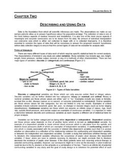
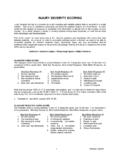
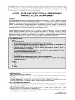
Transcription of cervical spine clearance 2009 - SurgicalCriticalCare.net
1 DISCLAIMER: These guidelines were prepared by the Department of Surgical Education, Orlando Regional Medical Center. They are intended to serve as a general statement regarding appropriate patient care practices based upon the available medical literature and clinical expertise at the time of development. They should not be considered to be accepted protocol or policy, nor are intended to replace clinical judgment or dictate care of individual patients. EVIDENCE DEFINITIONS Class I: Prospective randomized controlled trial. Class II: Prospective clinical study or retrospective analysis of reliable data. Includes observational, cohort, prevalence, or case control studies.
2 Class III: Retrospective study. Includes database or registry reviews, large series of case reports, expert opinion. Technology assessment: A technology study which does not lend itself to classification in the above-mentioned format. Devices are evaluated in terms of their accuracy, reliability, therapeutic potential, or cost effectiveness. LEVEL OF RECOMMENDATION DEFINITIONS Level 1: Convincingly justifiable based on available scientific information alone. Usually based on Class I data or strong Class II evidence if randomized testing is inappropriate. Conversely, low quality or contradictory Class I data may be insufficient to support a Level I recommendation.
3 Level 2: Reasonably justifiable based on available scientific evidence and strongly supported by expert opinion. Usually supported by Class II data or a preponderance of Class III evidence. Level 3: Supported by available data, but scientific evidence is lacking. Generally supported by Class III data. Useful for educational purposes and in guiding future clinical research. 1 Approved 4/18/2001 Revised 6/7/2005, 10/24/ 2009 cervical spine clearance SUMMARY cervical spine clearance following traumatic injury is an area of significant controversy. Patients often fall into one of two categories: those who are awake, alert, and able to participate in a clearance protocol and those who are obtunded and unable to facilitate clearance of their cervical spine .
4 A missed cervical spine injury can be devastating and may lead to chronic neck pain or even paralysis. Chronic use of cervical collars, however, is associated with the development of skin breakdown and ulceration. INTRODUCTION cervical spine injuries have been reported to occur in up to 3% of patients with major trauma and up to 10% of patients with serious head injury (1). The neurologic deficits that are a common consequence of these injuries can result in devastating disability for the patient as well as considerable economic and social burden for society. A missed injury can result in delayed treatment, instability, and possible quadriplegia; therefore, it is critical that physicians responsible for the initial evaluation and treatment of trauma patients be knowledgeable with regard to the methods for diagnosis of cervical spine injury.
5 Patients who are awake and alert can reliably be cleared of cervical spine pathology by painless clinical examination alone; patients who are obtunded, however, are much more difficult to assess and ultimately clear of cervical spine injury. clearance of the cervical spine should be based upon a thorough elucidation of the patient s history in addition to a complete physical examination. Many imaging modalities have been evaluated for the diagnosis of cervical spine injury. Plain radiographs (PR), static flexion & extension radiographs (F&E), computed tomography (CT), dynamic fluoroscopy (DF) and magnetic resonance imaging (MRI) have all been evaluated to determine their accuracy and usefulness in the diagnosis of cervical spine injury.
6 The choice of imaging modality to use initially is dependent upon the patient s history and physical examination as well as an assessment of the patient s risk for cervical spine injury. RECOMMENDATIONS Level 1 Awake and alert patients may be cleared by history and physical examination alone. Level 2 Computed tomography of the cervical spine from the occiput to T1 including axial, sagittal, and coronal images, should be utilized for cervical clearance . Due to the significant incidence of cervical collar-induced decubitus ulcers and other complications, cervical spine clearance should be performed within 72 hours of injury. Level 3 In the obtunded patient, computed tomography of the cervical spine is sufficient to allow clearance of the cervical spine .
7 2 Approved 4/18/2001 Revised 6/7/2005, 10/24/ 2009 LITERATURE REVIEW Clinical Examination vs. Plain Radiographs (PR) The National Emergency X-Radiography Utilization Study (NEXUS) was a prospective, observational study involving 21 centers across the United States that evaluated 34,069 stable patients with blunt trauma who were at risk for cervical spine injury (2). All patients were assessed using a straightforward, logical and easy to remember decision instrument that consisted of five clinical findings from the history and physical examination.
8 The presence of any one of these findings was considered to be evidence that the patient was at increased risk for a cervical spine injury and required radiographic evaluation. The NEXUS Clinical Criteria 1. Tenderness at the posterior midline of the cervical spine 2. Focal neurologic deficit 3. Decreased level of alertness 4. Evidence of intoxication 5. Clinically apparent pain that might distract the patient from the pain of a cervical spine injury The presence of any one of the above findings is considered to be clinical evidence that a patient is at increased risk for cervical spine injury and requires radiographic evaluation. A standard series of PR of the cervical spine (cross-table lateral view, anteroposterior view and open-mouth view of the odontoid) was obtained in all patients, unless CT or MRI of the entire cervical spine was performed because PR was impractical or impossible.
9 Other imaging studies could be ordered in addition to the standard series at the discretion of the treating physician. Diagnosis of cervical spine injury and determination of the type of injury was made according to the final interpretation of all the imaging studies. The decision instrument had a sensitivity of , a NPV of , a specificity of , and a PPV of for the identification of clinically significant cervical spine injuries. The Canadian C- spine Rule was derived from a prospective, observational study involving 10 centers across Canada that evaluated 8,924 alert and stable patients with blunt trauma who were at risk for cervical spine injury (3).
10 All patients were assessed for potential predictor variables of cervical spine injury. These variables consisted of 20 standardized clinical findings and 5 demographic variables from the patient s history and physical examination. Radiographic evaluation consisted of a standard series of PR of the cervical spine (cross-table lateral view, anteroposterior view and open-mouth view of the odontoid). Other imaging studies could be ordered at the discretion of the treating physician. Diagnosis of cervical spine injury and determination of the type of injury was made according to the final interpretation of all the imaging studies. Patients who did not undergo any radiographic evaluation underwent a structured 14-day proxy outcome measure by telephone.