Example: confidence
DOSE-EFFECT RELATIONSHIP
dissociation of the drug-receptor complex (the interaction is reversible). ... Although this curve resembles the graded dose-effect curve, remember that the graded curve expresses the relationship between intensity of effect with a change in the dose, whereas the quantal curve is a …
Tags:
Information
Domain:
Source:
Link to this page:
Documents from same domain
Structure and Mechanism of Alkaline Phosphatase
www.chem.uwec.eduSTRUCTURE AND MECHANISM OF ALKALINE PHOSPHATASE Joseph E. Coleman ... CONCLUSIONS ON STRUCTURE AND MECHANISM OF ALKALINE PHOSPHATASE DERIVED ... (66, 67). The amino acid sequences of the mammalian enzymes, derived from their cDNA sequences, can be fit into the primary structure of the bacterial enzyme, with the proper adjustments for some ...

Lecture 11 - Biosynthesis of Amino Acids
www.chem.uwec.edupentose phosphate pathway. The L–stereochemistry is enforced by transamination of α–keto acids Introduction. 5 Text ... 3–Phosphoglycerate dehydrogenase is inhibited by serine. 3. Regulation of Amino Acid Biosynthesis. 38 Text Example: Serine biosynthesis 3–Phosphoglycerate dehydrogenase 3. Regulation of Amino Acid

FTIR Analysis of Protein Structure
www.chem.uwec.eduB. Obtaining structural information about proteins using FTIR spectroscopy FTIR spectroscopy provides information about the secondary structure content of proteins, unlike X-ray crystallography and NMR spectroscopy which provide information about the tertiary structure. FTIR spectroscopy works by shining infrared radiation on a

Topic 7.2 INTRODUCTION TO DRUG DESIGN
www.chem.uwec.edu• Quick recovery times after surgery 1.2.9 Introducing chemically susceptible groups N CH 2 C O O MeO OMe H MeO (CH)5O C OMe O CH Me MeO OMe OMe OMe 1.2 Drug stability. ... Trojan horse tactic 1.3.1 Linking a biosynthetic building block 1.3 Drug targeting Non selective alkylating agent Toxic N Cl Cl H 3C Uracil Mustard H N HN O O N Cl Cl.

Chem 452 - Lecture 1 Introduction to Biochemistry Part 1
www.chem.uwec.eduevolved on earth Chem 452, Lecture 1 - Introduction to Biochemistry Biology is Varied and Complex 8 At the molecular level, living systems look remarkably similar. This similarity is a reflection of how life evolved on earth Chem 452, Lecture 1 - Introduction to Biochemistry Biology is Varied and Complex 8 At the molecular level, living systems

Chem 452 - Fall 2012 - Quiz 2 (Take Home, due Monday, …
www.chem.uwec.edub. What is the turnover number for Hexokinase under these conditions? # turnover number = k cat= V max [E]total = 186 µmol mL•s 3.0 nmol mL = 186x10−6 mol 3.0x10⁻⁹ mol mL =62,000/s c. What is the catalytic efficiency for Hexokinase under these conditions? # catalytic efficiency = k cat K M = 62,000/s 3.0x10⁻⁴ M =2.1 x 10⁸/(M•s) d. Does Hexokinase display “catalytic …
Chem 101: General Chemistry Lecture 1 - Matter ...
www.chem.uwec.edu3. Gases have mass. 4. Gases mix readily with one another. C. From these observations a simple model of matter was created which works not only with gases, but also with liquids and solids: 1. Matter is made up of tiny particles called molecules. 2. Molecules are the smallest division of matter that displays the chemical
Related documents

Cambridge International AS & A Level - GCE Guide
www.gceguide.com(d) Fig. 3.2 shows the oxygen dissociation curve for adult haemoglobin in a person who does not have sickle cell anaemia. 0 0 4 8 12 partial pressure of oxygen / kPa percentage oxygen saturation of haemoglobin 2 6 10 14 40 80 20 60 100 Fig. 3.2 Compared to Fig. 3.2, the oxygen dissociation curve for adult haemoglobin in a person with
3.2 Transport in Animals OCR ExamBuilder
www.calderlearningtrust.com(ii) Explain why it is important for the survival of the llama that the llama haemoglobin dissociation curve is to the left of the camel haemoglobin dissociation curve. [2]

QUANTITATIVE RT-PCR PROTOCOL (SYBR Green I)
schnablelab.plantgenomics.iastate.eduApr 01, 2007 · a. Check dissociation curve to make sure that every reaction just has single peak b. Check amplification curve to make sure there is no product formed for NTC (no contamination) c. Set up proper threshold to get the Ct values in the range of exponential amplification (usually 0.3 works fine) d.

Interpreting melt curves: An indicator, not a diagnosis
www.science.smith.eduFigure 3. Dissociation curve of CFTR exon 17b amplicon. Change in fluorescence with increasing temperature is measured. As the temperature is increased, the 2 strands of the amplicon separate to form single-stranded DNA, causing the fluorescent intercalating dye to dissociate from the DNA and stop fluorescing. DNA melting is a multi-state process

3- Oxygen Delivery Systems - Johns Hopkins HealthCare ...
www.johnshopkinssolutions.comOct 03, 2017 · Oxyhemoglobin Dissociation Curve KorupoluR GJ, NeedhamDM. Contemporary CriticalCare. 2009;6(9):1 ‐11 ...
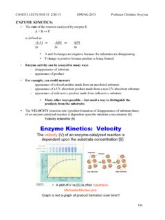
Enzyme Kinetics: Velocity - Purdue University
www.chem.purdue.eduThe Michaelis-Menten curve can be used to ESTIMATE V max and K M – although not exacting and we don’t use it. Determine the values by a different version of the equation. In 1934, Lineweaver and Burk devised a way to transform the hyperbolic plot into a linear plot. - Actual values for K M and V max can then be easily determined from the graph.

CELL CULTURE BASICS - Vanderbilt University
www.vanderbilt.eduMar 09, 2015 · Introduction Cell Culture Basics | 1 Purpose of the Handbook Cell Culture Basics Companion Handbook is a supplement to the Cell Culture Basics