Transcription of Examination Of The Abdomen
1 Examination Of The AbdomenPOM November 6, 2019 Charlie Goldberg, Exam 4 Elements: Observation, Auscultation, Percussion, Palpation Pelvic, male genital& male/female rectalexams all criticalparts of Abdomen exam covered later in the yearGI Review of Systems AnatomyUmbillicusSupra-Pubic AreaEpigastric AreaObservation & Draping Exposure Drapefor success expose what you need to see! Use sheet to cover lower half of body Good lighting, warmroom, table flat, handsat side, head resting on table +/-Feet flat on tableObservation (cont) Make note of : general shape contours symmetry color scars ? easiest to make observationsfrom footof bed. Examine from rightsideExamples of Abnormal Findings On ObservationObeseAscites (fluid), YellowEnlarged gall bladderUmbilical Hernia (Right with Valsalva)Auscultation Normal intestinalpropulsionof food (peristalsis) generates noise (Borborygmi) Listen(diaphragmof stethoscope) x 15-20 seconds in 4 quadrants Pay attention to: presence, quantity (normal ~ 2-5 seconds), & qualityof soundsAuscultation (cont) Clinical utility: Intestinal Obstruction: Increased frequencyearly ( rushes ) declinesin quantity, increase pitch ( tinkles ) stop Afterhandled (surgery) no functionor noise (ileus) w/normal recovery, noisereturns Infection of mucosa(gastroenteritis) increasedfrequency No findings pathognomonic Auscultation not helpfulin otherwise normal exam Clinical contextmost importantAuscultation (cont) Bruits.
2 Sounds of turbulentarterial flow atherosclerosis Relevant if: Unexplained hypertension, kidney disease, ischemic symptoms and risk factors Listen over: Renal arteries: several cm above umbilicus, either side rectus) Central Abdomen : celiac, SMA, IMA Iliac arteries: below umbilicusCeliac, SMA, IMAP ercussion Same principle as Lung Tappingover solidor liquidfilled structure dulltone; air filled tympanitic(resonant) Percussion what s beneathskin & bones : liver dull; air filled stomach tympanitic Abdomen not designed w/1st yrstudents in mind!-Key solid structures protected: liver & spleen by ribs; pancreas & kidneys deep in retro-peritoneum; bladder & uterus in pelvis-Central abdomenfilled w/intestines: freely moving promotes peristalsis, tolerates directtraumaPercussion Technique Stand on Right Middle fingerof non-percussing hand firmly against Abdomen Using floppy wristaction, hammer middle fingerof other handdown, aiming for last joint Percuss all 4 quadrants normal = s mix of dull and tympaniticPercussion Technique (cont) Liver span (6-12 cm): Startin chest, below nipple (mid-clavicular line) & move down tone changes from resonant (lung) to dull(liver) to resonant(intestines) Spleen small, located in hollow of ribs percussion over last intercostal space, anterior axillary line should normally be resonant dullness suggests splenomegaly Stomach tympaniticResonanceto percussionIf normal ( not enlarged)StomachPercussion To Detect Ascites.
3 Flank Dullness and Shifting Dullness Used to detect large amounts of pathological fluid (ascites) Intestineswill floatto surface Percussion can detect air-fluid interface Flank Dullness alone: Sensitivity: 84% Specificity: 59% Shifting Dullness: Sensitivity: 77% Specificity: 72% Intestines Ascites Simel. Rational Clinical Exam 2009. 65-69 Palpation Mostimportant structuresaren t palpable Warmyour hands Generally right handused (left placed on top or @ your side) Palpate using pads& edges of middle 3 fingers Gentle pressure, no sudden movements Think about what lives in areayou re examiningPalpation Technique First explore superficial aspect each quadrant(start R lower R upper Lupper Llower) Deeper palpationLiver Start R lower, moving uptowards R ribs Movehands a few cmup w/each palpation Push down(posterior) & then towardshead As approach ribs, palpatewhilepatient inspires deeply (diaphragm brings liver down towards hand) Might feel liver edgein normals(usually not)Palpation Technique (cont) Deeper Palpation (cont)Spleen Palpate towards left upperquadrant from midline & below -can use L hand to pull spleen towards youAorta (if RFs for aneurysm.)
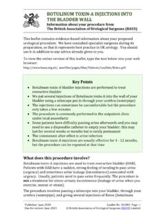
4 Age > 60, smoking) Above umbillicus, left of midline Push down (deep) w/palpating hand Remainder of Abdomen Uterus, bladder , other (rarely palpable) Evaluate painful areas last!Palpating to Detect fluid Wave (ascites) Examiner s right hand on patient s right Push quickly initiate a wave w/in ascites Receiving hand on Left identifies the wave A third hand dampens passage of wave through sub-cu fatSensitivity: 62% Specificity: 90%Simel. Rational Clinical Exam 2009. 65-69 Palpation/Percussion Of The Kidneys Kidneysare retroperitonealstructures, deep & protected by the ribs rarely palpable If markedly enlarged, may appreciate in lateral aspects Abdomen (rare) Assess for tenderness via posterior approach, tappingon backat Costo-Vertebral Angle if kidney infected (pyelonephritis), patient will have Tenderness (CVAT) Not done routinely: only in right clinical contextExposed Deep RetroperitoneumImage from U of Louisville, ICM CoursePut Findings Together Paint The Best PictureAbdominal exam techniques compliment each other!
5 Ascites Observe distention, bulging flanks Palpation no evidence of mass Palpation + fluid wave Enlarged liver(hepatomegaly) Percussion indicates extension of liver below diaphragm Palpation confirms location of lower edge (also detects contour, texture)Summary Of Skills Wash Hands Observe Abdomen (shape, contours, scars, color, etc) Auscultate Abdomen (bowel sounds, bruits) Percuss Abdomen (general; then liver & spleen) Palpate 4 quadrants Abdomen (superficial then deep) Assess for kidney area pain (CVAT) Wash HandsTime Target: < 10 Minutes