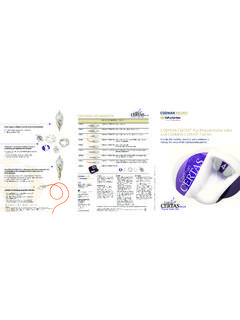
Transcription of Expert TN. Tibial Nail. - synthes.vo.llnwd.net
1 ExpertNailing SystemExpert TN. Tibial TechniqueInstruments and implants approved by the AO publication is not intended for distribution in the intensifier controlThis description alone does not provide sufficient background for direct use of depuy synthes products. Instruction by a surgeon experienced in handling these products is highly , Reprocessing, Care and MaintenanceFor general guidelines, function control and dismantling of multi-part instruments, as well as processing guidelines for implants, please contact your local sales representative or refer to: general information about reprocessing, care and maintenance of synthes reusable devices, instrument trays and cases, as well as processing of synthes non-sterile implants, please consult the Important Information leaflet (SE_023827) or refer to: Tibial nail Surgical Technique depuy synthes 1 TABLE OF CONTENTSINTRODUCTION Expert TN 2AO Principles 4 Indications 5 Cases 6 SURGICAL TECHNIQUE Preoperative Planning 8 Opening the Tibia 9 nail Insertion 19 Distal Locking 27 Proximal Locking 34 End Cap Insertion 54 Weight-bearing 56 Implant Removal 57 PRODUCT INFORMATION Implant Specifications 60 Implants 61 Instruments 68 Comparison Table 78 Handling Information 79 Modular Cases 80 Vario Case 83 Expert Tibial nail PROtect.
2 Why risk an infection? 85 Optional: Angular Stable Locking System (ASLS) 86 MRI INFORMATION 88 Stardrive T40 This patient has some synthes locking screws with hexalobular internal drive according to EN ISO 106640 mm 5 mm 10 mm 15 mm2 depuy synthes Expert Tibial nail Surgical TechniqueEXPERT TN Tibial NAILCOMPREHENSIVE SOLUTIONS Versatile proximal locking options: Three innovative locking options, in combination with can cellous bone lock-ing screws, increase the stability of the proximal fragment for proximal third fractures Two state of the art medio-lateral (ML) locking options enable primary compression or secondary controlled dynamizationCOMPREHENSIVE SOLUTIONS End caps: Securely lock the most proximal oblique locking screw to create a fixed-angle construct End cap prevents ingrowth of tissue and facilitates nail extraction Self-retaining Stardrive T40 recess for effortless end cap pick-up and ease of insertion Cannulated 0 mm end cap sits flush with nail 5 mm, 10 mm and 15 mm end caps extend nail height if nail is over insertedStardrive T25 This patient has some synthes locking screws with hexalobular internal drive according to EN ISO 10664 Expert Tibial nail Surgical Technique depuy synthes 3 COMPREHENSIVE SOLUTIONS All locking screws.
3 Double lead thread for more contact points for enhanced stability and ease of insertion Thread closer to screw head providing better bone purchase in the near cortex and improved stability Titanium alloy TAN* for improved me-chanical and fatigue properties Self-tapping blunt tip Self-retaining Stardrive T25 recess allows improved torque transmission and increased resistance to stripping relative to a hex recess and secure lock-ing screw pick-upCancellous bone locking screws: Indicated for the three proximal locking options of all Tibial nails diameters Dual core design for optimized purchase in cancellous bone Unicortical Lengths: 30 mm 90 mmStandard locking screws: Larger cross section for improved mechanical resistance B mm for B mm and B mm Tibial nails, lengths: 18 mm 80 mm B mm for B mm to B mm Tibial nails, lengths: 26 mm 100 mm* Titanium 6% aluminum 7% niobiumAdvanced nail design: Anatomic bend for ease of nail insertion Titanium alloy TAN* for improved mechanical and fatigue properties Cannulated nails (from B 8 mm to B 13 mm) for reamed or unreamed techniques, enabling nail insertion over guide wire The mm or mm ball tipped guide wires may be removed through the nail and insertion handle assembly (no exchange tube required).
4 Solid nails (from B 8 mm to B 10 mm) for unreamed techniqueAdvanced distal locking options: Distal oblique locking option to prevent soft tissue damage and increase stability of the distal fragment Two ML and one antero-posterior (AP) locking options for stability of the distal fragment14234 depuy synthes Expert Tibial nail Surgical TechniqueAO PRINCIPLESIn 1958, the AO formulated four basic principles, which have become the guidelines for internal fixation1, M ller ME, M Allg wer, R Schneider, H Willenegger. Manual of Internal Fixation. 3rd ed. Berlin Heidelberg New York: Springer. 19912 R edi TP, RE Buckley, CG Moran. AO Principles of Fracture Management. 2nd ed. Stuttgart, New York: Thieme. 2007 Anatomic reductionFracture reduction and fixation to restore anatomical , active mobilizationEarly and safe mobilization and rehabilitation of the injured part and the patient as a fixationFracture fixation providing abso-lute or relative stability, as required by the patient, the injury, and the personality of the of blood supplyPreservation of the blood supply to soft tissues and bone by gentle reduction techniques and careful Tibial nail Surgical Technique depuy synthes 5 INDICATIONSThe Expert Tibial nail is indicated for fractures in the tib-ial shaft as well as for metaphyseal and certain intraartic-ular fractures of the Tibial head and the pilon tibiale.
5 41-A 2 /A 3 All shaft fractures 43-A1/A2/A3 Combinations of these fracturesFor these indications the Expert Tibial nail should be used in combination with other implants (not shown in the illustrations): 41- C1/C2 43 - C1/C2 Note: The use of a cannulated Expert Tibial nail with a large diameter offering more stability associ-ated with the reamed technique is generally recom-mended for pseudarthroses, tumours, mal-unions and : ASLS, the Angular Stable Locking System, is indicated in cases where increased stability is needed in fractures closer to the metaphyseal area or in poor quality bone. For more details regarding the intramedullary fixator principle, please consult the ASLS technique guide ( ) and concept flyer ( ).Note: In cases with an increased risk of local bony infections, the Expert Tibial nail PROtect offers additional protection from bacterial colonization through local antibiotic prophylaxis.
6 For further information refer to page depuy synthes Expert Tibial nail Surgical TechniqueCASESF racture involving the proximal component Case 1 The use of the three locking screws in the proximal oblique locking options ensures optimal stabilization of the proximal fragment. The distal segment can be stabi-lized by using two ML locking options. Stability of the distal fragment can be enhanced by the use of a third locking screw in the AP hole. Shaft fractureCase 2 For simple shaft fractures, two proximal ML and two dis-tal ML locking screws are normally sufficient to stabilize the fracture. Secondary dynaminization is achieved by removing the proximal static locking involving the distal component Case 3 The use of four distal locking screws is sometimes neces-sary to achieve stabilization of the distal fragment. In many cases though, three locking screws placed in the most distal locking options are sufficient to stabilize the distal fragment.
7 Expert Tibial nail Surgical Technique depuy synthes 7 Case 1 Case 2 Case 3preoperativefollow-up (3 months after surgery)postoperativepreoperativepreoper ativefollow-up (1 month after surgery) follow-up (1 month after surgery) postoperativepostoperative8 depuy synthes Expert Tibial nail Surgical TechniquePREOPERATIVE PLANNINGUse the AO/ASIF Preoperative Planner Template for the Expert Tibial nail to estimate nail diameter and nail length. To estimate nail diameter, place the template on the AP or lateral X-ray of the uninjured tibia and mea-sure the diameter of the medullary canal at the narrow-est part that will contain the estimate nail length, place the template on the AP X-ray of the uninjured tibia and select the appropriate nail length based on patient anatomy. When selecting nail size, consider canal diameter, fracture pattern, patient anatomy and postoperative End Cap for Tibial NailsFor use only with the Original AO/ASIF System ofInstruments and Implants0102030405060708090100 Stratec Medical 2005 Printed in Switzerland SEK Subject to 8, 9, 10, 11, 12, 13 mm mm270 mm285 mm300 mm315 mm330 mm345 mm360 mm375 mm390 mm405 mm420 mm435 mm450 mm465 mmdynamicstatic0 mm513223715 10 5 0 mm 8 AP View Lateral ViewCancellous Bone Locking Screw mm (golden) mm30405060708090 Locking Screw mm (light green) mm26304050608010070 Locking Screw mm (dark blue) ( )7 mm 9 mm 10 mm 11 mm 12 mm 13 mmManufacturer: Stratec MedicalEimattstrasse 3CH-4436 Tibial nail Surgical Technique depuy synthes 9 OPENING THE TIBIA1 Position patientPosition the patient supine on the radiolucent table.
8 Ensure that the knee of the injured leg can be flexed at least 90 . Position the image intensifier such that visualisation of the tibia including the articular surface proximally and distally is possible in AP and lateral , the procedure can be performed on a fracture table with the leg placed in knee roller can be placed under the lower part of the thigh if it obstructs the view of the tibia plateau in AP depuy synthes Expert Tibial nail Surgical TechniqueOpening the Tibia2 Reduce fracturePerform closed reduction manually by axial traction under image intensifier. The use of the Large Distractor ( ) or Pinless Fixator in Vario Case ( ) may be appropriate in certain : The reduction can be temporarily fixed with reduction clamps. In epiphyseal fractures the condyles or the pilon tibiale are fixed first in order to enable the nail Tibial nail Surgical Technique depuy synthes 113 Confirm nail length and Radiographic Ruler for Expert Tibial NailThe required nail length must be determined after reduction of the lower leg the C-arm for an AP view of the distal tibia.
9 With long forceps, hold the ruler along the leg, parallel to and at the same level as the tibia. Adjust the ruler un-til the distal tip is at the level of the physeal scar or the desired nail insertion depth. Mark the skin at that site. Move the C-arm to the proximal tibia, replace the distal end of the ruler at the skin mark, and take an AP image of the proximal tibia. Read nail length directly from the ruler image, selecting the measurement at or just below the level of the anterior edge of the Tibial plateau. When using the large distractor, measure the distance from the inferior border of the distal pin to the superior border of the proximal pin to determine optimal nail length. Position the C-arm for an AP or lateral view of the tibia at the level of the isthmus. Hold the ruler over the tibia so that the diameter gauge is centered over the narrow-est part of the medullary canal.
10 Read the diameter measurement on the circular indicator that fills the : Compression or dynaminization must be taken into account when determining the nail length. A shorter nail should be chosen when active compres-sion is planned for the procedure. The dynamic locking option allows for 7 mm of : The ruler is not at the same level as the tibia. This affects the accuracy of the measurement, pro-viding only an estimated canal depuy synthes Expert Tibial nail Surgical TechniqueOpening the TibiaAlternativesDetermine the nail length by the above procedure on the uninjured leg or before draping (unsterile) or compare the length of two identical SynReam Reaming Rods B the radiographic ruler over the tibia so that the measuring edge is located over the isthmus. Select the nail diameter shown when the medullary canal/cortex transition is still visible on both sides of the marking.