Transcription of FACIAL LACERATIONS
1 FFAACCIIAALL LLAACCEERRAATTIIOONNSSThe face has several unique properties that dictate the choice of treat-ment after injury. This chapter describes basic principles for the treat-ment of FACIAL wounds as well as treatment recommendations forinjuries involving specific areas of the PPrrooppeerrttiieess ooff FFaacciiaall LLaacceerraattiioonnssCosmetic ConcernsAlthough most people do not want an unsightly scar anywhere on thebody, they are especially concerned about scars on their face. Thus, pri-mary closure, which usually results in the least noticeable scar, is thepreferred treatment for most FACIAL LACERATIONS . Fortunately, because ofthe laxity of FACIAL skin, most wounds can be repaired primarily unlessthey have significant tissue loss or tissue Blood Supply and CirculationThe skin of the face has a more abundant blood supply compared withother areas of the body. As a result, LACERATIONS on the face can be closedmore than 6 hours after injury (the usual time limit for closure of anacute laceration) without a high risk for subsequent wound long as the wound can be cleansed thoroughly, FACIAL lacerationsoften can be closed even the day after of the better blood supply, a wound that is closed primarilycan tolerate more tension on the suture line than is usually do nottake this principle to an extreme.
2 If there is significantblanching of the skin with the closure, you may not want to close thewound completely. In this instance, merely place a few sutures to closethe wound partially and thus decrease the size of the 16 KKEEYY FFIIGGUURREESS::Tissue flapLip anatomySuture bites: face vs. rest of bodySoft tissue loss146 Practical Plastic Surgery for NonsurgeonsIInniittiiaall CCaarreeThe initial care of a FACIAL wound is the same as the care applied to anywound. As explained in chapter 6, Evaluation of an Acute Wound, the wound needs to be cleansed fully and examined thoroughly. Allforeign material, blood, and necrotic tissue should be removed. Debride-ment of skin edges should be kept to a minimum, unless the tissue isobviously dead. Because of the excellent blood supply of the face,tissue that seems ischemic often the injured area with an antibacterial solution before closing thewound. Be careful: some solutions can cause injury to the eyes.
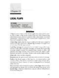
3 Tenpercent povidone iodine solution is commonly available and will notinjure the eyes. It also can be used safely on oral more thorough description of the administration of local anestheticsis found in chapter 3, Local Anesthesia. Below is a brief with epinephrine is the best choice of anesthetic with oneexception: when a flap is raised by the injury. In this case, it is best touse plain lidocaine in order not to diminish circulation to the flap in patient who fell through aglass window. A,Irregular forehead , The skin is separated from thedeep tissue layers of the forehead, creat-ing a skin flap with marginal blood not add epinephrine to the anestheticsolution; it will decrease circulation to theflap and may cause the tissue to die. C,One month after repair. All of the skin hassurvived and is healing without LACERATIONS 147 Bupivacaine is also acceptable. Add bicarbonate to decrease the pain of Local AnestheticFor smaller LACERATIONS (a few centimeters or less), it is often easiest toinject the anesthetic along the wound larger lacerationsor LACERATIONS around the edge of the lip (wherelocal injection can distort landmarks), a nerve block is usually Blocks on the FaceMental nerve block:lower lip, skin below the nerve block.
4 Upper lip, lateral nose, lower eyelid, nerve ChoiceNylon is the suture material of choice to close a skin wound on or other absorbable material should be used for you believe that the patient will not return for suture removal or ifthe patient is a child in whom suture removal is likely to be quite diffi-cult, chromic (absorbable material of choice) sutures can be used onfacial appropriate size of the suture is discussed in the sections describ-ing specific PlacementSutures on the face should be placed a little closer together than usu-ally recommended because of cosmetic concerns. The sutures shouldbe placed 1 2 mm from the skin edge and 3 mm apart to achieve bettertissue approximation. Exceptions are noted in specific you have access to magnifying glasses, use them. They help toachieve better tissue alignment because the magnification allows moreaccurate placement of the FACIAL LACERATIONS can be closed in one layer.
5 Exceptions will Practical Plastic Surgery for NonsurgeonsContinuous vs. Interrupted ClosureA laceration in which skin edges can be aligned easily and without ten-sion can be closed with either technique. Irregular LACERATIONS or LACERATIONS in which you are concerned aboutthe potential for infection should be closed in an interrupted fashionfor the following reasons:1. If, a few days after wound closure, a localized area starts to look in-fected, you can treat the infection without having to open the entirewound. Just remove a few sutures in the area that looks red, open theskin, and wash the wound with saline. This will allow the wound todrain and may allow the infection to resolve while keeping the resul-tant scar relatively small. The patient also should be given If the wound had been closed by placing the sutures in a continuousfashion, partial removal of the suture is not possible. If the woundlooks infected, the entire suture will need to be removed and thusthe entire wound will reopen.
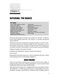
6 This results in a much larger bites: face vs. rest of the body. Sutures placed on the face should beapproximately 1 2 mm from the skin edge and approximately 3 mm apart. Thistechnique requires the use of small suture LACERATIONS 149 suture RemovalSutures should be removed after 5 7 days to minimize Instructions1. After the wound edges are sutured together, apply a small amountof antibiotic ointment over the suture line. Cover with a dry dressing can be removed on the following The area should be cleansed once or twice daily with gentle soapand water. The patient can shower and wash the face as usual on theday after the repair. 3. After cleansing, a small amount of antibiotic ointment or a petrola-tum type ointment should be applied over the suture line. If the pa-tient desires, dry gauze can be used to cover the area, although itusually is not necessary unless the patient is in a dirty FACIAL injuries cause the tissues to swell.
7 Be sure to warn your pa-tient that the face will be swollen for several days after injury. Tominimize swelling, instruct the patient to keep the head elevated atall times. When reclining, an extra pillow (or folded sheet) shouldbe placed under the The patient also should avoid bending and heavy lifting for severaldays after the injury because such activities promote FACIAL swelling. SSppeecciiffiicc WWoouunnddssLip LacerationsThe vermilion borderis the edge of the lip where the red part of the lipends and the white skin begins. It is vital to realign the vermilionborder meticulously to prevent a noticeable notched red part of the lip is the mucosal surface, which can be dividedinto two parts. The part of the lip that you see when the lips are barelyseparated is called the dry mucosabecause it feels dry to touch. Themucosal surface that lies against the teeth and appears and feels wet iscalled the wet mucosa.
8 These distinctions are important. Try to alignthe border between these two surfaces to prevent a relatively subtle,but noticeable make it easier to see the various borders, it is best to use a nerveblock for repair of lip LACERATIONS . If you are unable to do this, inject thelocal anesthetic a few millimeters away from the wound edge and waitlonger than usual (5 10 min) for the swelling from the injection to Practical Plastic Surgery for NonsurgeonsMucosal LACERATIONS The key to successful repair is to realign the wet-dry mucosal border,as explained above. Place the first stitch at the border between the wet and dry surfaces. Use absorbable sutures, 4-0, and try to sew the wet mucosa to wetmucosa and the dry mucosa to dry mucosa. It is important to evert the edges; use mattress sutures if necessary. Tie at least 4 or 5 knots in the sutures to prevent the sutures fromcoming undone because the patient unconsciously pulls at them withthe LACERATIONS that Cross the Vermilion Border The key to successful repair is to approximate the vermilion borderas well as possible.
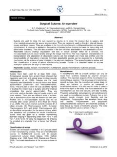
9 Align the red/white margin first. Place the initial suture just abovethe vermilion border in the white upper lip skin. Use a 5-0 or 6-0suture. If the stitch does not seem to be well-placed, remove it and try again. Place the remaining sutures in the lip skin (5-0 or 6-0) and lip mucosa(4-0 or 5-0). Be sure to evert the skin LacerationsIn full-thickness lip LACERATIONS , the outer skin, lip muscle, and mucosahave all been cut. Full-thickness lip LACERATIONS often look muscle retracts when cut, the lip wound looks larger and morecomplex than it is. Most of these wounds can be repaired repair is possible even if approximately one-fourth of theupper or lower lip is lost. Repair includes the following steps:Important anatomic landmarks of the LACERATIONS 1511. It often helps to place a gauze pad between the gums and lip to col-lect blood or other fluids. bleeding is significant:injection of lidocaine with epinephrineusually controls bleeding from the lip surface.
10 However, if thebleeding is coming from a cut artery, you may need to place a stitchin the artery. Use a 4-0 absorbable suture , and place a simple orfigure-of-eight suture at the site of bleeding. When you tie thesuture, the bleeding should the mucosa:repair the inner aspect of the lip first, as de-scribed above under Mucosal LACERATIONS . Use an absorbable 4-0suture, and try to evert the the woundwith saline again after the mucosa is closed tocleanse the the muscle:use an absorbable suture , 3-0 or 4-0, and placeone or two figure-of-eight sutures in the muscle. If you look care-fully at the wound edges, the muscle and musosa have a differentappearance and texture. Take care not to catch any mucosa in thestitches; if you do, you will cause a pucker in the the skin:as well as possible, align the vermilion border as de-scribed above. Remember to tie 4 or 5 knots in the lip sutures, whichoften come undone because the patient unconsciously pulls at them.