Transcription of GUIDE TO XRF BASICS
1 BRUKER ADVANCED X-RAY SOLUTIONS _____ GUIDE TO XRF BASICS _____ This GUIDE was first published in West Germany under the title Introduction to X-ray Fluorescence Analysis (XRF). 2000 - 2006 Bruker AXS GmbH, Karlruhe, West Germany. All rights reserved. Authors: Dr. Reinhold Schlotz, freelance writer and applications scientist, Bruker AXS Dr. Stefan Uhlig, International Sales Manager, Bruker AXS Bruker AXS GmbH stliche Rheinbr ckenstr. 49 D-76187 Karlsruhe Germany Tel: (+49) (721) 595-2888 Fax: (+49) (721) 595-4587 Email: Web: Bruker AXS Inc. 5465 East Cheryl Parkway Madison, WI 53711-5373 USA Tel: +1 (800) 234-XRAY Fax: +1 (608) 276-3006 Email: Web: Introduction to X-Ray Fluorescence (XRF) Table of Contents i Introduction to X-ray Fluorescence (XRF) Table of Contents 1. Fundamental Principles .. 1 Electromagnetic Radiation, Quanta.
2 1 The Origin of 2 Bohr's Atomic Model .. 2 Characteristic Radiation .. 3 Nomenclature .. 3 Generating the Characteristic Radiation .. 4 X-ray Tubes, Bremsspektrum .. 5 Tube Types, the Generator .. 6 Side-window Tubes .. 6 End-window Tubes .. 7 Generator .. 7 Excitation of Characteristic Radiation in Sample Material .. 8 Layer Thickness, Saturation 10 Secondary Enhancement .. 11 Tube-spectrum Scattering at the Sample 11 X-ray Detectors .. 12 Pulse Height Spectrum .. 12 Gas Proportional Counter .. 12 Scintillation 14 Pulse Height Analysis (PHA) .. 14 Pulse Height Distribution .. 14 The Counter 16 Diffraction in 17 17 18 X-ray Diffraction From a Crystal Lattice, Bragg's Equation .. 19 Reflections of Higher Orders .. 21 Crystal Types .. 22 Dispersion, Line Separation .. 24 Standard Types, Multilayers .. 24 Special Crystals.
3 26 Curved Crystals .. 31 2. Instrumentation .. 33 Multichannel 33 Scanners for Multichannel 34 Sequential 35 Incident Beam Components .. 37 Table of Contents Introduction to X-Ray Fluorescence (XRF) ii The End-window Tube and Generator .. 37 The Primary Beam 37 Sample Cups, the Cup 39 Emitted Beam Components .. 39 The Vacuum Seal .. 39 Collimator Masks .. 39 Collimators, the Soller Slit .. 39 The Crystal 40 The Flow Counter .. 40 The Sealed Proportional Counter .. 41 The Scintillation 42 Electronic Pulse Processing .. 42 The 42 Main Amplifier, Sine 42 Dead Time 42 Line-shift 44 3. Sample Preparation 45 Introduction .. 45 Preparation of Solid Samples .. 48 1 Metals .. 48 Pressed Pellets .. 48 Fused 49 Preparation of Liquid Samples .. 50 Preparation of Filter Samples .. 50 Appendix A Sources of Standard 51 Appendix B Supplementary Literature.
4 53 Books .. 53 53 55 Introduction to X-Ray Fluorescence (XRF) Fundamental Principles 1 1. Fundamental Principles Electromagnetic Radiation, Quanta From a physical point of view, X-rays are of the same nature as visible light. Visible light can be described as electromagnetic wave radiation whose variety of colors ( the colors of the rainbow) we interpret as different wavelengths. The wavelengths of electromagnetic radiation reach from the kilometer range of radio waves up to the picometer range (10-12 m) of hard gamma radiation (Table 1). Table 1: Energy and wavelength ranges of electromagnetic radiation Energy range (keV) Wavelength range Name < 10-7 cm to km Radio waves (short, medium, long waves) < 10-3 m to cm Microwaves < 10-3 m to mm Infra-red 380 to 750 nm Visible light 10 to 380 nm Ultra-violet 100 to 12 nm X-rays 10 5000 to nm Gamma radiation In the following text, the unit nanometer (nm = 10-9 m) is used for the wavelength, (= Lambda), and the unit kiloelectronvolts (keV) for energy, E.
5 Comment In literature the unit Angstr m ( ) is often stated for the wavelength: 1 = nm = 10-10 m The following relationship (conversion formula) exists between the units E (keV) and (nm): )( )(nmkeVE = or )( )(keVEnm= The X-ray fluorescence analysis records the following range of energy or wavelengths: E = 60 keV = nm Apart from the wave properties, light also has the properties of particles. This is expressed by the term photon . In the following we will be using the term quanta or X-ray quanta for this. The radiation intensity is the number of X-ray quanta that are emitted or measured per unit of time. We use the number of X-ray quanta measured per second, cps (= counts per second) or kcps (= kilocounts per second) as the unit of intensity. Fundamental Principles Introduction to X-Ray Fluorescence (XRF) 2 The Origin of X-rays Electromagnetic radiation can occur whenever electrically charged particles, particularly electrons, lose energy as a result of a change in their state of motion, upon deceleration, changing direction or moving to a lower energy level in the atomic shell.
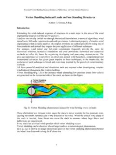
6 The deceleration of electrons and the transition from an energy level in the atomic shell to a lower one play an important part in the creation of X-rays in the field of X-ray analysis. To understand the processes in the atomic shell we must take a look at the Bohr's atomic model. Bohr's Atomic Model Bohr's atomic model describes the structure of an atom as an atomic nucleus surrounded by electron shells (Fig. 1): Fig. 1: Bohr's atomic model, shell model The positively charged nucleus is surrounded by electrons that move within defined areas ( shells ). The differences in the strength of the electrons bonds to the atomic nucleus are very clear depending on the area or level they occupy, they vary in their energy. When we talk about this we refer to energy levels or energy shells. This means that a clearly defined minimum amount of energy is required to release an electron of the innermost shell from the atom.
7 To release an electron of the second innermost shell from the atom, a clearly defined minimum amount of energy is required that is lower than that needed to release an innermost electron. An electron s bond within an atom is weaker the farther away it is from the atom s nucleus. The minimum amount of energy required to release an electron from the atom, and thus the energy with which it is bound in the atom, is also referred to as the binding energy of the electron in the atom. The binding energy of an electron in an atom is established mainly by determining the incident. It is for this reason that the term absorption edge is very often found in literature: Energy level = binding energy = absorption edge The individual shells are labeled with the letters K, L, M, N,.., the innermost shell being the K-shell, the second innermost the L-shell etc. The K-shell is occupied by 2 electrons.
8 The L-shell has three sub-Introduction to X-Ray Fluorescence (XRF) Fundamental Principles 3 levels and can contain up to 8 electrons. The M-shell has five sub-levels and can contain up to 18 electrons. Characteristic Radiation Every element is clearly defined by its atomic number Z in the periodic table of elements or by the number of its electrons in a neutral state. The binding energies or the energy levels in every element are different and characteristic for every element as a result of the varying number of electrons (negative charges) or the number Z of the positive charges in the atomic nucleus (= atomic number). If an electron of an inner shell is now separated from the atom by the irradiation of energy, an electron from a higher shell falls into this resultant hole which releases an amount of energy equivalent to the difference between the energy levels involved.
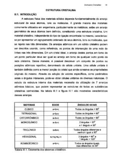
9 The energy being released can be either emitted in the form of an X-ray or transferred to another atomic shell electron (Auger effect). The probability of an X-ray resulting from this process is called the fluorescence yield . This depends on the element s atomic number and the shell in which the hole occurred. is very low for light elements (approx. 10-4 for boron) and almost reaches a value of 1 for the K-shell of heavier elements ( uranium). However, since the energy or wavelength of the X-ray is very characteristic for the element from which it is emitted; such radiation is called characteristic X-rays. This provides the basis for determining chemical elements with the aid of X-ray fluorescence analysis. Nomenclature The energy of an X-ray corresponds to the difference in energy of the energy levels concerned. K-radiation is the term given to the radiation released when replenishing the K-shell, L-radiation to that released when replenishing the L-shell etc.
10 (Fig. 2). Also needed for the full labeling of the emitted X-ray line is the information telling us which shell the electron filling the hole comes from. The Greek letters , , , .. are used for this with the numbering 1, 2, 3, .. to differentiate between the various shells and sub-levels. Fundamental Principles Introduction to X-Ray Fluorescence (XRF) 4 Fig. 2: X-ray line labeling Examples: K 1 Electron from sub-level LIII to the K-shell K 2 Electron from sublevel LII to the K-shell K 1,2 if neither line is resolved by the spectrometer K 1 Electron from sublevel M to the K-shell L 1 Electron from sublevel M to the L-shell Generating the Characteristic Radiation The purpose of X-ray fluorescence is to determine chemical elements both qualitatively and quantitatively by measuring their characteristic radiation. To do this, the chemical elements in a sample must be caused emit X-rays.