Transcription of LOCAL FLAPS - Practical Plastic Surgery
1 LLOOCCAALL FFLLAAPPSSDD eeffiinniittiioonnssAflapis a piece of tissue with a blood supply that can be used to coveran open wound. A flap can be created from skin with its underlyingsubcutaneous tissue, fascia, or muscle, either individually or in somecombination. Depending on the reconstructive requirements, evenbone can be included in a flapimplies that the tissue is adjacent to the open wound inneed of coverage, whereas in a distant flap , the tissue is brought froman area away from the open wound. LOCAL flap coverageof a wound is the next higher rung up the recon-structive ladder after a skin graft.
2 Examples of wounds that require flapcoverage include wounds with exposed bone, tendon, or other vitalstructure and large wounds over a flexion crease, for which a split-thickness skin graft or secondary closure would result in tight site:where the flap site:the open wound/soft tissue defect in need of :the blood supply of the flap ( , its arterial inflow andvenous outflow). The pedicle varies from a wide bridge of tissue (skin,subcutaneous tissue, muscle, or some combination) to an isolatedartery and vein.
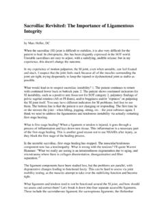
3 Most LOCAL FLAPS can be classified as either (1) skin FLAPS , which are skinand subcutaneous tissue with or without the underlying fascia, or (2)muscle FLAPS , which are created from a muscle with or without the at-tached overlying 13 KKEEYY FFIIGGUURREESS::Axial flap noting pedicleRhomboid flapRandom flap notingRotation flappedicle and 3:1 ratioV-Y advancement flap112 Practical Plastic Surgery for NonsurgeonsSSkkiinn FFllaappssA portion of skin and subcutaneous tissue and, when possible, the under-lying fascia (the thin layer of connective tissue overlying muscle that hasan excellent vascular supply) is moved to fill the defect.
4 This movement oftissue results in a new defect at the donor site. Often the donor site can beclosed primarily, but sometimes a skin graft is FLAPS are classified as either axialor random. The classification isbased on the blood FlapsThe circulation of an axial flap is supplied by specific, identifiableblood vessels. Careful anatomic study has identified several donorsites with a single artery and vein responsible for circulation to a par-ticular area of skin. Examples include the volar forearm skin suppliedby the radial artery and skin on the back supplied by the circumflexscapular artery (a branch of the thoracodorsal artery).
5 Circulation based on specific vessels results in a highly reliable bloodsupply and a reliable flap . You can be confident that unless there is aninjury to the vessels, the majority of the flap tissue should survive in itsnew position. Axial flap . Note that the blood supply comes from an identifiable vessel. As aresult, the pedicle can be quite thin, which makes transferring the flap to itsnew site an easier FLAPS 113An axial flap can be completely detached from all surrounding tissue aslong as it remains connected to its supplying blood vessels.
6 These ves-sels serve as the pedicle. The thin pedicle allows axial FLAPS to be easilypositioned to fill the wound defect (unlike the random flap [see below]). The difficulty with an axial flap is locating the blood vessels. You mustbe very careful not to injure the vessels when creating the flap . The nec-essary technical expertise is beyond the realm of most providers with-out reconstructive surgical training. Thus, no specific axial skin flapsare discussed in this FlapsCirculation to a random flap is provided in a diffuse fashion throughtiny vascular connections from the pedicle into the flap .
7 The pediclemust be bulky to increase the number of vascular connections. Themore vascular connections, the better the circulation to the flap . Thebetter the circulation to the flap , the better its general, a random flap does not have as reliable a blood supply asan axial flap . Nonetheless, the relative ease of creating random flapsmakes them useful almost anywhere on the body. The circulation andthus the reliability of the flap can be increased by delaying the flapbefore final transfer. Delay ProcedureBefore the flap is created, the tissue gets its blood supply via all of thesurrounding skin and underlying tissue attachments.
8 When the flap iscreated, the circulation to the flap comes only from the skin flap . The blood supply comes diffusely from the remaining skinattachment, which serves as the pedicle. For optimal circulation and flap sur-vival, the flap should be designed so that the length is no more than three timesthe Practical Plastic Surgery for NonsurgeonsThe purpose of the delay procedure is to enable the pedicle to assumeits role as the main source of circulation before the flap is moved to itsnew position. This goal is obtained by making some of the incisionsneeded to create the flap but not separating the flap from the underly-ing tissues.
9 The flap is not moved to its new position; instead, the skinedges are sutured together total blood supplied to the flap initially decreases when the inci-sions are made. This decrease promotes opening of new vascular chan-nels between the pedicle and flap . Thus, more blood will flow into theflap through the pedicle than if the delay procedure had not been the flap before final transfer allows more confidence in theviability of the flap . Wait about 7 10 days after the delay procedurebefore moving the flap to the recipient ffoorr CCrreeaattiinngg RRaannddoomm LLooccaall FFllaappssWhen creating a random LOCAL skin flap , you take advantage of the rel-atively loose, excess skin in the vicinity of the skin defect.
10 Randomflaps require less technical expertise than axial FLAPS . Because they canbe quite useful for covering an open wound, several types of randomflaps are discussed in detail InformationRandom flap procedures often can be done under LOCAL anesthesia ifthe area ( flap plus defect) is not too large (< 8 10 cm). For larger areas,general anesthesia probably will be sure to clean the wound thoroughly before creating and placing the a scrub brush or the flat part of a scalpel to scrape away the top layerof granulation tissue from the wound.