Example: bachelor of science
MITRAL VALVE STENOSIS - asecho.org
Assessment of Mitral Stenosis • Mitral valve area measurement – > 1.5 cm2-Mild – 1.1 to 1.5 cm2-Moderate – < or = 1.0 cm2-Severe Quantification of Mitral Valve Area • Direct Planimetry • Pressure Half-Time • Continuity / PISA
Tags:
Information
Domain:
Source:
Link to this page:
Documents from same domain

Aortic Regurgitation: Etiology and Echo …
asecho.orgAortic Regurgitation: Etiology and Echo Quantification Martin G. Keane, MD, FASE Professor of Medicine Lewis Katz School of Medicine at Temple University

Recommendations for Noninvasive Evaluation of …
asecho.orgASE GUIDELINES AND STANDARDS Recommendations for Noninvasive Evaluation of Native Valvular Regurgitation A Report from the American Society of Echocardiography
Recommendations for the Evaluation of Left …
asecho.orgASE/EACVI GUIDELINES AND STANDARDS Recommendations for the Evaluation of Left Ventricular Diastolic Function by Echocardiography: …

Echocardiography in Pulmonary Arterial …
asecho.orgSTATE-OF-THE-ART REVIEWARTICLES Echocardiography in Pulmonary Arterial Hypertension: from Diagnosis to Prognosis Eduardo Bossone, MD, PhD, Antonello D’Andrea, MD, PhD, Michele D’Alto, MD, Rodolfo Citro, MD,
Recommendations for Evaluation of Prosthetic …
asecho.orgGUIDELINES AND STANDARDS Recommendations for Evaluation of Prosthetic Valves With Echocardiography and Doppler Ultrasound A Report From the American Society of Echocardiography’s Guidelines and Standards

2017 Coding and Reimbursement Newsletter. …
asecho.org{D0700524.DOCX / 1 } o If you perform moderate sedation in conjunction with the TEE or other procedures that you provide, use the following new CPT
Left Atrial Appendage Closure Devices - asecho.org
asecho.org10/10/2017 1 Muhamed Sarić MD, PhD, MPA Director of Noninvasive Cardiology | Echo Lab Associate Professor of Medicine Left Atrial Appendage Closure Devices

Guidelines for Cardiac Sonographer Education ...
asecho.orgcardiac sonographer. Specific goals of this revision include the following: • Define the role of the cardiac sonographer • Provide recommendations for educational path-

Multimodality Imaging Guidelines of Patients with ...
asecho.orgASE GUIDELINES AND STANDARDS Multimodality Imaging Guidelines of Patients with Transposition of the Great Arteries: A Report from the American Society of Echocardiography Developed

EAE/ASE Recommendations for Image Acquisition …
asecho.orgTABLE OF CONTENTS 1. Introduction 4 2. Instrumentation 4 a. Fully Sampled Matrix-Array Transducers 4 3. Data Acquisition 5 a. Challenges with 3DE Ac-
Related documents
Transcatheter Heart Valve Procedures - UHCprovider.com
www.uhcprovider.comDiagnosis of severe calcific native aortic valve stenosis as indicated by one of the following: o Mean aortic valve gradient ≥40 mmHg; or o Peak aortic jet velocity ≥4.0 m/s; or o 2Aortic valve area of ≤ 0.8 cm Individual is symptomatic (New York Heart Association [NYHA] class II or greater) and symptoms are due to aortic valve stenosis
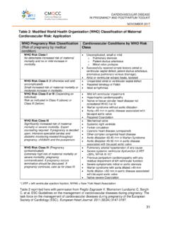
Table 2: Modified World Health Organization (WHO ...
www.cmqcc.org• Severe systemic ventricular dysfunction (LVEF <30%, NYHA III-IV)* • Previous peripartum cardiomyopathy with any residual impairment of left ventricular function • Severe symptomatic mitral or aortic stenosis • Marfan syndrome with aorta dilated >45 mm • Aortic dilation >50 mm in aortic disease associated with bicuspid aortic valve
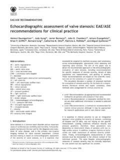
Echocardiographic assessment of valve stenosis: EAE/ASE ...
www.escardio.orgAortic stenosis Echocardiography has become the standard means for evaluation of aortic stenosis (AS) severity. Cardiac ... or severe (extensive thickening and increased echogenicity with a prominent acoustic shadow). The degree of valve calcification is a …

Transcatheter aortic valve implantation (TAVI) procedure
www.kch.nhs.ukcalled aortic stenosis. As a result, the heart cannot easily pump blood through the narrowed valve. This causes symptoms including breathlessness and chest pain. Some people may ... • 5% risk of a moderate to severe leaky valve • 5-10% risk of a slow heart rate requiring a permanent pacemaker • 3% risk of stroke

Aortic Regurgitation - asecho.org
www.asecho.orgInitially Asymptomatic Aortic Regurgitation Events: Heart Failure, New AFib, Cardiac Death Cardiac event rate (%) Years Mild AR Moderate AR Severe AR Detaint D, et al: JACC 2008; 1:1 P<0.0001 5±3 21±8 27±5 34±6 47±7 63±8 Quantitative Doppler