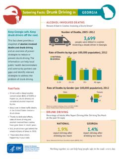
Transcription of Myeloid Malignancies
1 Myeloid Malignancies John Howard, Administrator, World Trade Center Health Program February 1, 2014 Executive Summary Beginning on February 1, 2014, the World Trade Center (WTC) Health Program will consider blood or bone marrow disorders of the Myeloid line to be slow-growing blood cancers. Accordingly, they will be considered WTC-related health conditions, making them available for WTC Health Program medical treatment services for eligible members. These cancers had been considered non-malignant by the Administrator because they were referred to as pre-leukemic hematopoietic disorders in the medical literature. Recent scientific advances, however, characterize these pre-leukemic Myeloid neoplasms as slow-growing blood cancers, and authoritative scientific sources now consider them to be malignant Myeloid neoplasms.
2 After receiving a request from the WTC Clinical Centers of Excellence to review certain Myeloid disorders in terms of their status as Malignancies ,1 the WTC Health Program has determined that, in addition to types of leukemias, these Myeloid Malignancies are eligible for coverage by the WTC Health Program as WTC-related health The group of Myeloid Malignancies includes the following health conditions: (1) Myelodysplastic Syndromes (MDSs); (2) Myeloproliferative neoplasms (MPNs); (3) Myelodysplastic/myeloproliferative neoplasms (MDS/MPN); and (4) Myeloid Malignancies associated with eosinophilia and abnormalities of growth factor receptors derived from platelets or 1 Letter to Drs. Dori Reissman and John Halpin of the WTC Health Program from World Trade Center Clinical Centers of Excellent Principal Investigators dated December 16, 2013 (on file at WTC Health Program).
3 2 See 42. (Table 1). 3 acute Myeloid leukemia (AML) remains eligible for coverage as a WTC-related health condition because it is already included in the List of WTC-Related Health Conditions. See 42 (Table 1). Myeloid Malignancies February 1, 2014 Page 2 of 9 I. Introduction In December of 2013 the WTC Clinical Centers of Excellence (CCEs) requested that the WTC Health Program review certain Myeloid disorders in terms of their status as MDS is one type of a group of Myeloid Malignancies . Therefore, based on the CCEs request, the Administrator reviewed the available scientific literature and authoritative disease classification sources pertaining to the malignancy of Myeloid neoplasms. The term Myeloid includes all cells belonging to the granulocyte ( , neutrophil, eosinophil, basophil), monocyte/macrophage, erythroid, megakaryocyte, and mast cell lineages.
4 Myeloid Malignancies are clonal diseases of hematopoietic stem or progenitor These Malignancies can be present in the bone marrow and peripheral blood. They result from genetic and epigenetic alterations that perturb key processes such as self-renewal, proliferation and impaired ,6 Some Myeloid disorders, such as the Myeloid leukemias, have long been considered malignant while other Myeloid disorders have been considered non-malignant or pre- leukemia blood disorders which may become malignant over time. However, recent scientific findings indicate that these pre- leukemia blood disorders are actually forms of slow-growing blood cancers. 7,8 Based on the morphology, cytochemistry, immunophenotype, genetics, and clinical features of Myeloid disorders, the World Health Organization (WHO) categorizes Myeloid Malignancies into five primary types: (1) acute Myeloid leukemia ; (2) myelodysplastic syndromes (MDS); (3) myeloproliferative neoplasms (MPN); (4) myelodysplastic and myeloproliferative (MDS/MPN) neoplasms; and (5) Myeloid neoplasms associated with eosinophilia and abnormalities of growth factor receptors derived from platelets or fibroblasts.
5 The types and subtypes of Myeloid Malignancies are identified in Table 1 in the Appendix. 4 Murati A, Brecqueville M, Devillier R, Mozziconacci M, Gelsi-Boyer V, Birnbaum D [2012]. Myeloid Malignancies : mutations, models and management. BMC Cancer 12:304-325. 5 Shih AH, Abdel-Wahab O, Patel JP, Levine RL [2012]. The role of mutations in epigenetic regulators in Myeloid Malignancies . Nat Rev Cancer 12(9):599-612. 6 Ntziachristos P, Mullenders J, Trimarchi T, Aifantis I [2013]. Mechanisms of epigenetic regulation of leukemia onset and progression. Adv Immunol 117:1-38. 7 Ma X [2012]. Epidemiology of myelodysplastic syndromes. Am J Med. 125(7 Suppl): S2 S5. 8 Tefferi A, Vardiman JW [2008]. Classification and diagnosis of myleoproliferative neoplasms: the World Health Organization criteria and point-of-care diagnostic algorithms.
6 leukemia 22:14-22. Myeloid Malignancies February 1, 2014 Page 3 of 9 II. Risk Factors A. Myelodysplastic Syndrome and Myeloproliferative Neoplasms The primary risk factor for MDS is age. The majority of secondary MDS cases occur after treatment for other cancers with radiation therapy or chemotherapy that employs alkylating agents or topoisomerase inhibitors. In addition, several environmental and/or occupational exposures have been associated with increased rates of MDS or cytogenetic abnormalities associated with MDS including pesticides9, benzene10, organic solvents11, semi-metals12, and inorganic dusts13. Studies of occupations identified increased incidence of MPN among poultry workers, commercial pressmen, petroleum refinery workers, agricultural workers, cooks/waiters and clerks.
7 Studies of associations with exposure to chemicals such as benzene, petroleum solvents, hair dyes, and pesticides have produced inconsistent B. Myeloid Malignancies Other Than AML, MDS and MPN Information on associations of environmental or occupational exposures for other Myeloid Malignancies was not found in a Medline search of the relevant medical literature. 9 Vundinti BR, Kerketta L, Jijina F, Ghosh K [2009]. Cytogenetic study of Myelodysplastic syndrome from India. Indian J Med Res 130:155-159. 10 Corey SJ, Minden MD, Barber DL, et al. [2007]. Myelodysplastic syndromes: the complexity of stem-cell diseases. Nat Rev Cancer 7:118-129. 11 Rigolin GM, Cuneo A, Roberti MG, Bardi A, Bigoni R, Piva N, Minotto C, Agostini P, De Angeli C, Del Senno L, Panedda R, Castoldi G [1998].
8 Exposure to myelotoxic agents and myelodysplasia: case-control study and correlation with clinicobiological findings. Br J Haematol 103:189-197. 12 West RR, Stafford DA, White AD, Bowen DT, Padua RA [2000]. Blood 95:2093-2097. 13 Vardiman JW, Thiele J, Arber DA, Brunning RD, Borowitz MJ, Porwit A, Harris NL, Le Beau MM, Hellstrom-Lindberg E, Tefferi A, Bloomfield CD [2009]. The 2008 revision of the World Health Organization (WHO) classification of Myeloid neoplasms and acute leukemia : rationale and important changes. Blood 114:937-951. 14 Reviewed by: Anderson LA, Duncombe AS, Hughes M, Mills ME, Wilson JC, McMullin MF [2012]. Environmental, lifestyle, and familial/ethnic factors associated with myeloproliferative neoplasms. Am J Hematol 87(2):175-182. Myeloid Malignancies February 1, 2014 Page 4 of 9 III.
9 Clinical and Pathologic Features The clinical and pathologic features of Myeloid Malignancies vary according to type. acute Myeloid leukemia . AML results from the clonal expansion of Myeloid blasts in the peripheral blood, bone marrow or other tissue. It is caused when either the Myeloid stem cells produce abnormal myeloblasts which do not become healthy white blood cells or too many Myeloid stem cells become abnormal red blood cells or platelets. As a result, leukemic blasts, or immature cell forms, accumulate in the bone marrow, peripheral blood, and occasionally in other tissues, and the production of normal red blood cells, platelets, and mature granulocytes are reduced a variable amount. The increased production of malignant cells, along with a reduction in these mature elements, results in a variety of systemic consequences including anemia, bleeding, and an increased risk of Myelodysplastic syndromes.
10 MDSs are a spectrum of bone marrow failure disorders that share the common pathologic feature of cytological dysplasia. They progress to acute Myeloid leukemia (AML) in about 30% of patients. MDSs are classified according to features of cellular morphology, cellular and molecular genetics, immunophenotyping, etiology, and clinical presentation. The seven subtypes of MDSs are listed in Table 1 in the Appendix. The morphological classification of MDSs is largely based on the percent of myeloblasts in the bone marrow and blood, the type and degree of Myeloid dysplasia, and the presence of ring sideroblasts. MDSs remain among the most challenging of the Myeloid Malignancies to diagnose and classify, particularly in cases in which the blast percentage is not increased in the peripheral blood or bone Myeloproliferative Neoplasms.