Transcription of 食道裂孔のOmentalHerniationの1例 - jrs.or.jp
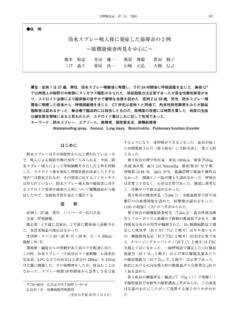
1 36 1 1998 67.. Omental Herniation 1 . 1 1 2 1 . 48 X CT MRI . omental herniation omental herniation .. Omental herniaton Esophageal hiatus 153 cm 82 kg .. omental herniation 1 CT MRI .. X .. Fig 1 . 48 CT . 100 HU .. Fig 2 . 8 9 MRI high intensity 9 . Fig 3 . Fig 1 Chest X-ray on admission, showing the large, sharply defined, retrocardiac mass. 1 099 0404 3 .. 2 078 8510 4 5 . 1 . 9 3 18 . 68 36 1 1998 . Fig 2 Chest CT scan showing an encapsulated retro- Fig 3 MRI coronary scan and schema, showing a ret- cardiac low density mass upper scan The mass ex- rocardiac high intensity mass extending into the ab- tended into the upper abdomen with no clear capsule domen through the right side of the esophageal hia- lower scan tus. CT MRI. omental herniation .. Fig 4 . CT MRI . fat density .. omental herniation.
2 Fig 4 Celiac arteriogram showing an artery supplying . the mass arrows .. 3 1 2 4 2 3 5 . Table 1 .. Omental Herniation 1 69. Table 1 Case reports of paraoesophageal omental herniation Preoperative No. Year Author Age sex Symtom Therapy diagnosis 1 1966 Pomerantz 50. M none unknown operation 2 1977 Rohlfing 50. M crampy postprandial pain POH operation 3 1982 Irisawa 37. M none lipoma operation 4 1988 Tamura 56. M none POH or lipoma operation 5 1990 Lee 76. M none POH follow-up 6 1997 Saijo 48. F none POH follow-up POH : paraoesophageal omental herniation .. paraoesophageal omental hernition5 1 Pomerantz RM, Twigg HL : Intrathracic omental herniation. J Thorac Cardiovasc Surg 1996 ; 52 : CT fat den- 735 736. sity 4 2 Rohlfing BM, Korobkin M, Hall AD : Computed To- CT 1 mogaphy of Intrathoracic Omental Herniation and Other Mediastinal Fatty Masses.
3 J Comput Assist 2 . Tomogr 1977 ; 1 : 181 183. CT 3 . 3 . 4 . 1 .. 1982 ; 41 : 560 565. 4 . 4 Omental Herniation 1 . CT MRI 1988 ; 26 : 1010 1014. Lee 5 CT MRI 5 Lee MJ, Breathanach E : Case Report : CT and MRI. Findings in Paraoesophageal Omental Herniation. Clinic Radiology 1990 ; 42 : 207 209. 6 DeMartine WJ, House AJS : Paratial Bochdalek's Bochdalek herniation. Chest 1980 ; 77 : 702 704. 7 . Morgagni . 1974 ; 12 : 6 13 Morgagni . 403 407.. 8 Morgagni . 14 . 4 1978 ; 40 : 557 561. 15 . 9 . Morgagni 1981 ; 34 : 325. 10 . Rohlfing 2 Morgagni 1 CT. 1984 ; 32 : 588 596. 11 Gossios KJ, Tatsis CK, Lykouri A, et al : Omental Morgagni Herniation Through the Foramen of Morgagni Daiagnosis with Computed Tomography. Chest LEE 5 1991 ; 100 : 1469 1470. 12 . X CT. Morgagni 1 .. 1992 ; 53 : 1330 1333. 13.
4 70 36 1 1998 . Morgagni 1 Surg 1966 ; 52 : 461 468. 1993 ; 54 : 3026 3029. 15 Bentley G, Lister J : Retrosternal hernia. Surgery 14 Comer TP, Clagett OT : Surgical treatment of her- 1965 ; 57 : 567 575. nia of the foramen Morgagni. Thorac Cardiovasc Abstract A Case of Paraesophageal Omental Herniation Yasuaki Saijo*, Hajime Honda*, Yutaka Nishigaki** and Tadataka Noro*. *Department of Cardiology, Engaru Kousei General Hosital, Engal-cho, Hokkaido, Japan **First Department of Internal Medicine, Asahikawa Medical College, Asahikawa city, Hokkaido, Japan A 48-year-old woman underwent routine chest roentgenography and a mass shadow was seen in the poste- rior mediastinum. CT, MRI and celiac arteriography were performed, and paraesophageal omental herniation was diagnosed. Paraesophageal omental herniation is uncommon, and there have been no reports cases with complica- tions.
5 Therefore, this case is being followed-up carefully.