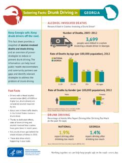
Transcription of Ophthalmology Procedures Manual
1 Ophthalmology Procedures Manual September 2005 TABLE OF CONTENTS Chapter Page 1 OVERVIEW OF 1-1 1-1 General Overview of 1-2 Integrated Survey Information System (ISIS) .. 1-2 2 EQUIPMENT/SUPPLIES/MATERIALS .. 2-1 Ophthalmology Equipment and Supplies .. 2-1 Nonconsumables (Instruments and Equipment).. 2-1 Supplies (Consumables).. 2-2 Equipment Description, Setup and Operating 2-2 Humphrey Matrix visual Field 2-2 Canon CR6-45NM Ophthalmic Digital Imaging System and Canon EOS 10D Digital 2-10 3 PROTOCOL .. 3-1 Eligibility 3-1 Safety Exclusion Questions Completed in 3-2 Pre-examination Procedures .
2 3-5 visual Field Test (Frequency Doubling Technology Perimetry).. 3-6 Setting Up ISIS and FDT screens .. 3-6 SP Positioning for the visual Field Test .. 3-9 Starting the visual Field 3-11 Saving Files to the ISIS Database .. 3-12 FDT Section Status .. 3-14 Procedure for Testing One Eye Only .. 3-15 Problems With Importing 3-16 Digital Fundus Photography .. 3-21 ISIS Screen for Retinal Images .. 3-22 Positioning for Retinal Imaging .. 3-23 Achieving Maximum Pupil 3-23 Explanation of Retinal Imaging .. 3-23 iii TABLE OF CONTENTS (continued) Chapter Page Camera Alignment and Imaging Procedures .. 3-24 Pupil Size and External Camera 3-25 Internal Eye Alignment Macula Image .. 3-26 Internal Eye Alignment Optic Nerve Image .. 3-27 Labeling the Images .. 3-27 Criteria for Taking Repeat Images.
3 3-29 Ignore/Image Not 3-32 Imaging Procedures for Challenging Circumstances .. 3-33 Data Acquisition 3-37 Completing the 3-40 Retinal Imaging Section Status .. 3-41 Storing and Shipping 3-42 4 QUALITY 4-1 4-1 4-1 Initial Training .. 4-1 Followup Training Prior to Main 4-2 Training of New 4-2 Equipment and Room Setup 4-2 Quality Control Log-on Box .. 4-2 Utilities Menu to Select Quality 4-3 Quality Control Log-on Box .. 4-4 Daily Quality Control 4-4 Weekly Quality Control Checks .. 4-5 Start of 4-6 Review of Images by 4-7 Observational 4-7 Site Visit Report Form .. 4-8 5 REFERRALS AND REPORT OF FINDINGS .. 5-1 Report of Findings.
4 5-1 Early Reporting of 5-1 Preliminary 5-2 iv TABLE OF CONTENTS (continued) Chapter Page Detailed Grading .. 5-2 Final Report of Findings .. 5-2 List of Appendixes Appendix A Step-By-Step Procedures and Scripts .. A-1 B Step-By-Step Procedures and Scripts - Spanish .. B-1 C Equipment Diagrams .. C-1 List of Tables Table 2-1 Troubleshooting problems with the Humphrey Matrix visual Field Instrument .. 2-9 2-2 Settings for the Canon EOS 10D camera 2-13 List of Figures Figure 2-1 Humphrey Matrix visual Field Instrument (examiner side).. 2-3 2-2 Humphrey Matrix visual Field Instrument (SP side) .. 2-3 2-3 SP Response 2-4 2-4 2-4 2-5 Power connector.
5 2-4 2-1 Canon CR6-45NM ophthalmic digital imaging 2-10 2-2 Canon EOS 10D digital 2-10 v TABLE OF CONTENTS (continued) List of Figures (continued) Figure Page 3-1 Safety exclusion observations from vision 3-1 3-2 Safety exclusion questions completed in Ophthalmology .. 3-2 3-3 Exclusion due to eye 3-3 3-4 Component Status due to eye infection exclusion .. 3-3 3-5 Exclusion due to eye patch-both 3-4 3-6 Component Status due to eye patch 3-4 3-7 FDT Data Capture 3-6 3-8 FDT Main Menu Screen .. 3-7 3-9 FDT View Patient Screen .. 3-7 3-10 FDT Enter New Patient 3-8 3-11 FDT View Patient Screen .. 3-8 3-12 FDT Testing Screen .. 3-9 3-13 FDT Eye Positioning .. 3-10 3-14 FDT SP Video Screen 3-10 3-15 Recall Test Screen-Saving files .. 3-13 3-16 Recall Test Screen Selecting format .. 3-13 3-17 Recall Test Screen Files saved successfully.
6 3-14 3-18 ISIS Data Capture Screen .. 3-14 3-19 FDT Selection Status Screen .. 3-15 3-20 Proceed without 3-16 3-21 DOB and ID does not 3-17 vi TABLE OF CONTENTS (continued) List of Figures (continued) Figure Page 3-22 Import failed .. 3-17 3-23 Files not found .. 3-18 3-24 More than two sets of 3-19 3-25 Data already 3-19 3-26 Imported two sets of data already .. 3-20 3-27 FDT Section Status for one file imported .. 3-20 3-28 Diagram of optic fields for 3-21 3-29 Retinal Images Screen .. 3-22 3-30 Drop-down menu for estimating pupil size in mm .. 3-26 3-31 Retinal Images Screen with drop-down menu .. 3-29 3-32 Images required by protocol .. 3-30 3-33 Image display for additional images .. 3-31 3-34 Missing entries message.
7 3-31 3-35 Ignore image .. 3-32 3-36 Image not 3-33 3-37 Drop-down menu for pupil size setting .. 3-35 3-38 ISIS screen for changing pupil setting .. 3-35 3-39 Apply pupil size setting to all subsequent images .. 3-36 3-40 ISIS screen for changing DA setting .. 3-37 3-41 Apply DA setting to all subsequent 3-37 3-42 Data acquisition screen .. 3-38 vii TABLE OF CONTENTS (continued) List of Figures (continued) Figure Page 3-43 Rating difficulty of taking 3-39 3-44 Rating quality of the 3-40 3-45 Retinal Images Section Status 3-41 3-46 Burn DVD 3-43 3-47 Burn DVD message .. 3-43 3-48 DVD burn status bar .. 3-44 3-49 Assign air bills assigned 3-44 3-50 Assigned air bills- not assigned.
8 3-45 4-1 Quality control reminder message box .. 4-3 4-2 Utilities menu to select quality control .. 4-3 4-3 Quality control 4-4 4-4 Daily QC checks .. 4-5 4-5 Weekly QC 4-6 4-6 Start of stand QC 4-6 4-7 Site Visit Report 4-8 viii 1. OVERVIEW OF Ophthalmology Background The leading causes of visual impairment in the United States are primarily age-related eye diseases including cataracts, diabetic retinopathy, glaucoma, and age-related macular degeneration. More than million Americans aged 40 years and older are either blind or are visually impaired. Although it is believed that half of all blindness can be prevented, the number of people with blindness continues to increase in the United States. Unfortunately, scant data exist for national estimates and trends, and current estimates are based on data that are 25 years old and not nationally representative.
9 Glaucoma is the leading cause of irreversible blindness and is a prevalent disease associated with aging. Although glaucoma can usually be controlled by early detection and treatment, half of the people with glaucoma are not diagnosed, and glaucoma is still the number one blinding disease among African Americans. Diabetic retinopathy is the leading cause of new cases of blindness among adults aged 20-74 years. It can affect almost anyone with diabetes and contributes to both individual and societal burden. With the growing epidemic of diabetes and demographic changes in the American society, vision loss and eye diseases due to diabetes will be a growing major public health problem. Efficacious and cost-effective strategies to detect and timely treat diabetic retinopathy are available, but among people with diabetes ocular eye examination is received by only about two-thirds of the persons for whom the exam is recommended and varies significantly across health care settings.
10 Age-related macular degeneration (AMD) is the leading cause of visual impairment and blindness in the among people aged 65 years or older. The frequency of AMD is expected to increase as the population lives longer. Population-based estimates of the prevalence and severity of AMD will help in allocating resources as treatment modalities become available. 1-1 General Overview of Procedures Prior to the Ophthalmology testing, the SP will complete the NHANES Vision examination component which includes visual acuity and objective refraction for SPs aged 8 years and older in addition to a near vision exam on SPs 50 years and older. The visual acuity and objective refraction tests in the current vision component must be completed prior to completing the examinations in the Ophthalmology component because the flash from the retinal imaging may affect the visual acuity results.