Transcription of Optimized Immunohistochemistry Workflow …
1 1 Colley & Stead 110531* Corresponding Author E-mail: This e-mail address can be publishedOptimized Immunohistochemistry Workflow Facilitated by New Dako Autostainer Link 48 Software Elizabeth C. Colley, MLT, ART and Ronald H. Stead, PhD, FRCPath* Molecular and Cellular Pathology Laboratory, Gamma-Dynacare and Holburn Biomedical Corporation 1100 Bennett Road, Bowmanville, Ontario, Canada, L1C 3K5 Telephone: 905 697 4711 or 905 623 1484 x220 Facsimile: 905 697 1415 or 905 623 6702 2 Colley & Stead 110531 AbstractAutomated Immunohistochemistry systems, including instruments and reagents, are used by many histopathology laboratories. Dako currently supplies one such instrument, the Autostainer Link 48. This has a capacity of 48 slides per batch/run and uses a pipetting arm to aspirate and dispense reagents.
2 Until recently, the production software (version ) resulted in staining times of up to hours, excluding antigen retrieval and ancillary steps. However, a new software version ( ) has just been introduced, and was evaluated in the authors laboratory. Using software version , to stain over 40 slides, resulted in run times of about hours compared with over hours using software version software. The new software version also reduced buffer usage and generated less waste than the previous version. Including dewaxing, antigen retrieval (in Dako s PT Link), staining on the Autostainer Link 48, counterstaining, dehydration, clearing and coverslipping, the Dako system processes 96 slides in approximately hours and 144 slides in about hours, which is greater throughput than competitive platforms.
3 Moreover, the new software version significantly reduces hands-on time per slide compared to other capacity of currently available IHC instruments varies from about 30 to 60 slides. The most commonly used systems are from Ventana, Leica and Dako (3-5), all of which provide dedicated reagent sets for use with their instrument platforms. While other manufacturers also supply instrument and reagent packages, most of these function similar to the Dako system (3). With previous versions of the operating software, the Dako Autostainer took longer to complete a full staining run, not including antigen retrieval in the PT Link instrument (6). Even though the Dako system could stain 48 slides at once, compared with 30 for Ventana and Leica, it was generally considered to have a lower throughput, which would be a disadvantage for a busy diagnostic recently, the Dako system s longer staining times have been due to software scheduling issues for the pipetting arm not to the staining method per se.
4 To address this issue and improve run times, Dako has developed new software (version ). In the current study, the authors compared the processing capacity and other features of the version software running on an Autostainer Link 48 instrument, in parallel with an Autostainer Link 48 instrument running the version software. The study was conducted in the Gamma-Dyncare Molecular and Cellular Pathology Laboratory in Bowmanville, Ontario, Canada. Introduction: Immunohistochemistry (IHC) is a key diagnostic tool in tissue pathology (1). The number of antibodies available for application to routine formalin-fixed, paraffin-embedded (FFPE) tissue sections is increasing rapidly. While some laboratories still employ manual methods to perform immunohistochemical stains, many institutions increasingly use automated staining platforms.
5 Some of these platforms are open and can employ any available primary antibody or detection system; and others are closed , requiring at least the use of detection systems provided by the instrument manufacturer. Over the years, automated staining instruments have made use of various mechanisms to dispense immunohistochemical reagents. For example, capillary gap technology was used to draw reagent between two directly opposed slides ( , patient and control) (2). However, most systems have relied upon robotic pipetting mechanisms, sometimes with ancillary delivery systems for wash buffers and other bulk fluids. Accordingly, the time taken for any IHC instrument to perform a full staining run depends not only on incubation times of individual reagents, but also on the time it takes for robotic arms to move from one slide station to another, or from reagent wells to slides.
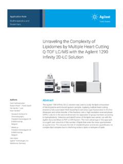
6 Hence, the actual throughput rate for any automated IHC instrument is based on both the total time required for the instrument to perform a full staining run and on the numbers of slides stained. Optimized Immunohistochemistry Workflow Facilitated by New Dako Autostainer Link 48 Software 3 Colley & Stead 110531 Results Comparison of software and new software version (version ) for the Autostainer Link 48 instrument resulted in 30% faster run times (up to two hours time saved) compared with version Run Time ComparisonRun Time (min)501-910-1920-29 Number of Slides 1: Run time comparisons for a range of slide batchesRed bars indicate version software; blue bars indicate version software. The light blue circles above each bar represent the mean predicted run time for each group (batch size as indicated on the x-axis; n = 4 or 5 per group).
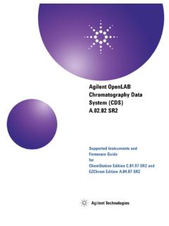
7 The maximum variation was seen in the group of 30-39 slides stained using version software (mean (n): (4)). Run time is clearly improved with the new software, especially with larger slide 2: Examples of results from pairs of slides stained using both versions of of results from pairs of slides stained with versions (A, C, and E) and (B, D, and F) software. Equivalent results were consistently attained. A & B: submucosal nodule stained for actin; C & D: normal small bowel stained for vimentin; and E & F: granular cell tumour stained with S100. IHC was performed on formalin-fixed, paraffin-embedded (FFPE) sections using Dako FLEX Ready-to-Use Primary Antibodies and the EnVision FLEX detection with full batches of 48 slides (Figure 1). Predicted run times were also improved.
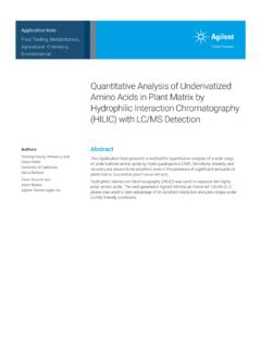
8 Importantly, faster run times did not affect overall IHC staining quality at least not for the 39 primary antibodies ABCDEF4 Colley & Stead 110531and over 1,000 slides evaluated in this study. There was, however, some indication that staining was slightly stronger using version software, with no increases in background staining. Examples of the paired staining results are shown in Figure 2. In no instance was the difference in staining intensity greater then (on an arbitrary scale 0 to 3+). More importantly, any differences in staining intensity would not affect the interpretation of the stain. Consistency of Staining Consistency of staining using version software was very good. Sample microscopic views of slides from the second of four full runs are shown in Figure 3. The quality of staining is clearly excellent, and is equivalent to results using version software.
9 The consistency is at least as good as results seen with competitive Additional Studies Reduced scanning timeThe formal study of scan times confirmed initial observations: scan times are shorter using version software than with version As might be expected, scan times were longer when more slides were introduced using version software. However, a very interesting finding was that the scan time with version software was relatively consistent regardless of the number of slides stained: approximately two minutes per run. This is shorter than the minimum scan time using version software with only a few slides on the instrument (approximately three minutes), and much shorter than the circa six minutes required for a full batch. Although this results only in a four-minute saving, there is an added benefit in those instances in which low or expired reagents are detected during the scan, necessitating a second scan after adding or replenishing those solutions.
10 Instruments from Ventana and Leica, recently evaluated in the authors laboratory (unpublished data). A few slides did exhibit staining variability. However, this was in tissue located very close to the label end of the slide. Other minor staining defects were rarely observed in these slides or in the paired sections stained with other antibodies. These appeared to be either roughly circular or angular, with reduced staining, and are likely due to small bubbles in one or more reagents. The reader should be aware that such artifacts are not restricted to the Dako Autostainer platform; these have been seen in all other systems tested in the authors laboratory over the past few years. In some cases the other instruments exhibited significantly more of these 3: Example of consistency of staining using new software.