Transcription of Pathophysiology and Pathogenesis of Osteomyelitis
1 1 Pathophysiology and Pathogenesis of Osteomyelitis Mayank Roy1, Jeremy S. Somerson1, Kevin G. Kerr2 and Jonathan L. Conroy2 1 University of Texas Health Science Centre, San Antonio, Texas 2 Harrogate District Hospital, North Yorkshire 1 USA 2UK 1. Introduction The term Osteomyelitis encompasses a broad group of infectious diseases characterized by infection of the bone and/or bone marrow. The Pathogenesis of these diseases can follow acute, subacute or chronic courses and involves a range of contributory host and pathogen factors. A commonly used aetiological classification distinguishes between three types of Osteomyelitis : acute or chronic haematogenous disease seeded by organisms in the bloodstream, local spread from a contiguous source of infection and secondary Osteomyelitis related to vascular insufficiency.
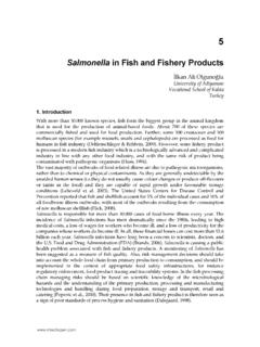
2 Acute haematogenous Osteomyelitis Acute haematogenous Osteomyelitis refers to infection of bone resulting from bacteria in the bloodstream. This is seen most often in children, with initial infection thought to occur in the richly vascularised metaphyseal region (Gutierrez, 2005). Children are thought to experience frequent episodes of bacteraemia, often with no apparent symptoms, leading to seeding and development of Osteomyelitis (Conrad, 2010). The Pathogenesis of this process has been theoretically described. Inoculation of the metaphyseal vessels occurs at the transition point from the arteriolar vessels to the venous sinusoids, slowing blood flow and increasing vascular turbulence (Jansson et al., 2009). These sites of turbulence may be predisposed to bacterial infection by providing an opportunity for local invasion (Fig.)
3 1). Although rarely seen in developed countries, haematogenous Osteomyelitis may take on a chronic course within bone if left untreated. Sequelae of this devastating condition may include chronic sinuses with exposed bone, loss of structural integrity and growth disturbances (Beckles et al., 2010). Local trauma to bone in the setting of bacteraemia may also be a contributing factor. Animal studies have shown significantly increased rates of haematogenous Osteomyelitis when direct injury to bone was combined with intravenous bacterial seeding. (Kabak et al., 1999; Morrissy & Haynes, 1989). A recent series of 450 cases of acute haematogenous Osteomyelitis found the rate of preceding blunt trauma to be 63% (Labbe et al., 2010). Further research is needed to elucidate the role of trauma in this condition.
4 Osteomyelitis 4 Fig. 1. Schematic drawing showing the vascular supply to the physis. The callout represents a detailed view of the physis. The red arrow indicates an area of transition. These transitional zones show increased turbulence and allow for local invasion. (Image used with permission from Dr. Kaye Wilkins) Vertebral Osteomyelitis Osteomyelitis involving the spine is also most commonly caused by haematogenous seeding of bacteria into the vertebrae (Tay et al., 2002). The Pathophysiology of this condition reflects the unique vascular structures of the spine. The venous anatomy of the spine, originally investigated for its role in cancer metastasis, allows retrograde flow from the pelvic venous plexus due a lack of valvular structures, providing an opportunity for haematogenous deposition of bacteria (Batson, 1967).
5 Fine arteriolar structures surrounding the vertebral end plate may also represent a location at which bacteria can become trapped (Wiley & Trueta, 1959). Infections are predominantly localized to the lumbar and thoracic spine, with significantly less frequent involvement of the cervical spine (Beronius et al., 2001). In children, a markedly different disease process has been observed in infections of the spine. Blood vessels in the paediatric spine pass through the physeal cartilage and terminate within the intervertebral disc, allowing for seeding of infection from the osseous vasculature (Tay, et al., 2002). This results in a direct extension of infection into the disc that is not seen in adult patients (Fig. 2). For this reason, this condition is referred to by some authors as paediatric discitis rather than Osteomyelitis .
6 Osteomyelitis secondary to contiguous infection In adult patients , the majority of Osteomyelitis cases are due to inoculation from contiguous infection. Sources can include direct contamination at a site of injury, iatrogenic contamination at the time of an invasive procedure, or invasive infection from surrounding soft tissue. The epidemiology of contiguous infection Osteomyelitis is biphasic, with young patients suffering trauma and related surgery and older patients suffering decubitus ulcers Pathophysiology and Pathogenesis of Osteomyelitis 5 Fig. 2. MRI scan showing disc space infection. The lack of normal disc signal at the circled segment (black arrow) represents infection. This type of spinal infection is seen more commonly in children. (Image used with permission from Dr. Kaye Wilkins). (Mader et al.)
7 , 1999). Chronic infection often results, with clinical courses complicated by loss of bone structural integrity and soft tissue envelope disturbance. The progression of disease in localized Osteomyelitis is characterized by a cycle of microbial invasion, vascular disruption, necrosis and sequestration. The host inflammatory response, discussed in detail below, results in obstruction of small vessels due to coagulopathy and oedema. As a result of this, cortical bone undergoes necrosis and is detached from surrounding live bone, creating an area known as a sequestrum. This provides a fertile environment for further bacterial invasion and progression continues. Simultaneously, induction of bone begins at the intact periosteum, forming a layer of viable osseous tissue around the site of infection known as involucrum.
8 This mechanism is thought to result from an inflammatory reaction of the periosteum. Osteomyelitis of the diabetic foot represents a common form of localized infection. Aetiological factors have been thought to include peripheral neuropathy with associated superficial ulceration and peripheral vascular disease. However, a large recent study of risk factors for Osteomyelitis in 1666 diabetic patients found no association of Osteomyelitis with either peripheral neuropathy or vascular disease (Lavery et al., 2009). History and physical examination findings associated with increased relative risk for Osteomyelitis in this study Osteomyelitis 6 included a previous history of foot ulceration prior to enrolment, the presence of multiple foot wounds or wounds that penetrated deep to bone or joint.
9 This supports prior literature suggesting that clinical ability to probe bone directly in a diabetic ulcer is diagnostic of underlying Osteomyelitis (Grayson et al., 1995). 2. Host factors The Pathogenesis of Osteomyelitis is a complex process involving interactions between a host and an infectious agent. The host s inflammatory response to a pathogen can further the physical spread of disease by clearing space in bone. Predisposing genetic differences in immune function are increasingly seen as an aetiological factor in some cases of Osteomyelitis . Acquired factors such as diseases causing immune or vascular compromise and implantation of foreign materials are frequently involved in the disease process as well. Inflammatory response to infection The unique demarcated environment of Osteomyelitis results in a high-grade local inflammatory host response with systemic effects ranging from minimal to severe.
10 The initial host response to infection of bone is characterized by a local increase in proinflammatory cytokines. Involvement of human monocyte cells in this process has been well-described. When presented with Staphylococcus aureus cells or bacterial cell wall components such as peptidoglycan (PepG) or lipopolysaccharide (LPS), monocytes secrete large amounts of interleukin 1-beta (IL-1beta), IL-6, IL-8, tumour necrosis factor alpha (TNF-alpha) and macrophage inflammatory protein 1-alpha (MIP-1alpha) (Fullilove et al., 2000; Klosterhalfen et al., 1996; Wang et al., 2000). This has been confirmed in an in vivo animal model demonstrating up-regulation of cytokines following intravenous infusion of PepG and LPS (Ruud et al., 2007). Matrix metalloproteases, a zinc-dependent group of endopeptidases, have been proposed as a key element of bone loss in Osteomyelitis .