Transcription of Plasmodium malariae
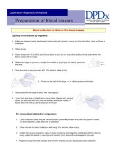
1 Laboratory diagnosis of malaria Plasmodium malariae Basic guidelines A. Capillary blood should be obtained by fingerstick, or venous blood should be obtained by venipuncture. B. Blood smears, at least two thick and two thin, should be prepared as soon as possible after collection. Delay in preparation of the smears can result in changes in parasite morphol-ogy and staining characteristics. C. Sch ffner s dots can be demonstrated in Giemsa stain, which is preferred to Wright or Wright-Giemsa stains. In P. malariae infections, red blood cells (rbcs) are normal or smaller than normal (3/4 ) in size. 1. Rings P. malariae rings have sturdy cytoplasm and a large chromatin dot. Ring in a thick blood smear . Rings in thin blood smears. 2. Trophozoites P. malariae trophozoites have compact cytoplasm and a large chromatin dot.
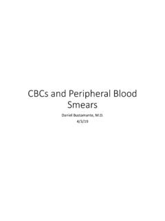
2 Occasional band forms and/or basket forms with coarse, dark-brown pigment can be seen. Trophozoite in a thick blood smear . Band-form trophozoites in thin blood smears. Laboratory diagnosis of malaria Plasmodium malariae Trophozoite in a thick smear . Band-form trophozoites in thin blood smears. Basket-form trophozoite in a thin Basket-form trophozoites in a thin smear . Trophozoites in thin blood smears. These images show variations on the basket-form. Laboratory diagnosis of malaria Plasmodium malariae 3. Schizonts P. malariae schizonts have 6 to 12 merozoites with large nuclei, clustered around a mass of coarse, dark-brown pigment. Merozoites can occasionally be arranged as a rosette pattern. Schizont in a thick blood smear . Schizont in thick blood smears. Note the classic rosette appearance of the merozoites.
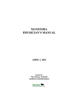
3 Schizonts in thin blood smears. The schizont on the left has the appearance of a rosette pattern. Schizont in a thin blood smear ; note the rosette pattern of the merozoites. Schizonts in thin blood smears Schizont in a thin blood smear ; another resembling a rosette. Laboratory diagnosis of malaria Plasmodium malariae 4. Gametocytes P. malariae gametocytes are round to oval with scattered brown pigment; they may almost fill the infected red blood cell. Gametocytes in thick blood smears. Gametocyte in a thin blood smear . Gametocyte in thin blood smears. Gametocytes in thin blood smears.